. 1998 Jun 15;509 ( Pt 3)(Pt 3):833-45.
doi: 10.1111/j.1469-7793.1998.833bm.x.

Kainate receptor-mediated inhibition of presynaptic Ca2+ influx and EPSP in area CA1 of the rat hippocampus

Affiliations

Kainate receptor-mediated inhibition of presynaptic Ca2+ influx and EPSP in area CA1 of the rat hippocampus

H Kamiya et al. J Physiol. .

Abstract

1. The effect of a low concentration (1 microM) of kainate (kainic acid; KA) on presynaptic calcium (Ca2+) influx at the Schaffer collateral-commissural (SCC) synapse was examined in rat hippocampal slices. 2. Following selective loading of the presynaptic terminals with the fluorescent Ca2+ indicator rhod-2 AM, transient increases in the presynaptic Ca2+ concentration (pre[Ca2+]t) and field excitatory postsynaptic potentials (EPSPs) evoked by electrical stimulation of the SCC pathway were recorded simultaneously. 3. Bath application of 1 microM KA reversibly suppressed field EPSPs and pre[Ca2+]t to 37.7 +/- 4.0 % and 72.9 +/- 2.4 % of control, respectively. Excitatory postsynaptic currents (EPSCs) recorded with the use of the whole-cell patch-clamp technique were also suppressed by 1 microM KA to 42.6 +/- 6.3 % of control. A quantitative analysis of the decreases in pre[Ca2+]t and the amplitude of field EPSP during KA application suggests that KA inhibits transmission primarily by reducing the pre[Ca2+]t. 4. Consistent with a presynaptic site for these effects, paired-pulse facilitation (PPF) was enhanced by 1 microM KA. 5. A substantial KA-induced suppression of NMDA receptor-mediated EPSPs was detected when AMPA receptors were blocked by the AMPA receptor-selective antagonist GYKI 52466 (100 microM). 6. The suppressive effect of KA on field EPSPs and pre[Ca2+]t was antagonized by the KA antagonist NS-102 (10 microM). 7. These results suggest that the presynaptic inhibitory action of KA at the hippocampal CA1 synapse is primarily due to the inhibition of Ca2+ influx into the presynaptic terminals.


Figures

Figure 1
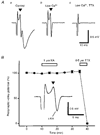
Figure 1. Selective loading of presynaptic terminals with the Ca2+ indicator rhod-2 AM
A, schematic diagram showing the experimental arrangement. Membrane-permeable rhod-2 AM (0.2 mM) was pressure ejected into the stratum radiatum, resulting in selective loading of the presynaptic terminals through the Schaffer collateral-commissural (SCC) pathway. Fluorescence from the shaded area, which has a diameter of about 100 μm and is about 500 μm away from the ejection site, was recorded with a single photodiode. B, time courses of the presynaptic Ca2+ transient (pre[Ca2+]t, upper trace) and the field EPSP (lower trace) evoked by a single stimulus to the SCC pathway. Ca2+ signals were measured as relative fluorescence changes (ΔF/F), where F is the resting fluorescence level and ΔF, the peak amplitude of fluorescence change caused by the stimulus. C, effects of a mixture of AMPA and NMDA antagonists (10 μM CNQX and 25 μM D-AP5) on the Ca2+ transient (upper traces) and field EPSP (lower traces). Two traces obtained before (control) and during application of the drugs are superimposed. D, effects of Ca2+-free solution containing 1 mM EGTA on pre[Ca2+]t and field EPSP.
Figure 2
Figure 2. Rhod-2 fluorescence transients and field EPSPs induced by paired-pulse stimulation
A, superimposed traces of fluorescence transients (upper traces) and field EPSPs (lower traces) in response to single and paired-pulse stimulations (50 ms interstimulus interval). B, the fluorescence transient and field EPSP produced by the second stimulus (dotted traces) were extracted by subtracting the traces evoked by single stimulation from the paired responses, and are shown together with those evoked by single stimulation.
Figure 3
Figure 3. Reversible inhibition of pre[Ca2+]t and field EPSPs by KA
A, representative records of pre[Ca2+]t and field EPSPs observed before, during and after washout of 1 μM kainate (KA). B, time courses of the KA effects on pre[Ca2+]t and field EPSPs. The relative amplitudes of pre[Ca2+]tF/F;○) and field EPSPs (•) evoked every 5 min were plotted as a function of time (mean ± s.e.m., n = 6). The mean of the first three values before drug application was expressed as 100 %. KA (1 μM) and a mixture of the AMPA receptor antagonist CNQX (10 μM) and the NMDA antagonist D-AP5 (25 μM) were applied during the period as indicated.
Figure 4
Figure 4. Relationship between pre[Ca2+]t and field EPSPs
A, linear plots of the pre[Ca2+]tF/F) and the field EPSP measured during application of KA (1 μM, n = 6; •) or domoate (0.2 μM, n = 6; ▴), or during replacement with low Ca2+ solution (containing 1.2 mM Ca2+ and 2.5 mM Mg2+, n = 6; ○) for 10 min. Data are normalized to control values (×) obtained before each manipulation. B, log-log plots of the data shown in A. The linear regression line for low Ca2+ data (○) was plotted as a dotted line, and the slope (m) was estimated to be 2.8. The corresponding power-law fit is also shown in A as a dotted curve.
Figure 5
Figure 5. Enhancement of paired-pulse facilitation (PPF) by KA
A, specimen records of field EPSPs (mean of 10 consecutive responses) evoked by paired stimuli (50 ms interval) in control solution (upper trace) and during KA application (1 μM, 10 min; lower trace). B, superimposed traces of the scaled field EPSPs before and during KA application. The EPSP component of the field potentials was extracted by subtraction of presynaptic fibre volleys and stimulus artifacts recorded in the presence of 10 μM CNQX and 25 μM D-AP5. The first EPSP amplitude in the presence of KA was normalized to that in the control record. These traces clearly demonstrate that PPF is larger during KA application. C, percentage facilitation of the second EPSPs (100 % indicates that the second response is twice as large as the first response in this figure) in the absence (control, ▪) and presence of 1 μM KA (□). * Difference statistically significant (n = 6, t test, P < 0.05).
Figure 6
Figure 6. Effects of KA on presynaptic fibre potentials
A, field potentials shown on a faster time scale. In the control solution (a), the field potentials (mean of 10 consecutive responses) consist of stimulus artifacts, presynaptic fibre volley potentials (▾) and the following EPSP components. Replacement with low Ca2+ solution (0.1 mM Ca2+ and 3.6 mM Mg2+) abolished field EPSPs, leaving only artifacts and presynaptic volleys (b). The presynaptic volley potentials were abolished by the further addition of 0.5 μM tetrodotoxin (TTX; c). B, plot of presynaptic volley amplitude as a function of time (n = 6). Field potentials were recorded in the low Ca2+ solution, and KA (1 μM) and TTX (0.5 μM) were applied during the period as indicated by the open bars. Insets show the specimen records of presynaptic fibre volley potentials recorded in the absence and presence of 1 μM KA.
Figure 7
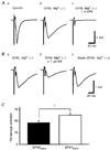
Figure 7. Effects of KA in the presence of the AMPA-selective antagonist GYKI 52466
A, recordings of field EPSPs mediated by NMDA receptor activation (field EPSPNMDA). To facilitate the recording of the NMDA component of EPSPs in isolation, a modified Mg2+-free solution containing glycine (10 μM) and GYKI 52466 (100 μM) was used. Switching to the Mg2+-free solutions resulted in isolation of the field EPSPNMDA of slower time course (b) than in normal solution (a). Further addition of D-AP5 (25 μM) abolished field EPSPNMDA, leaving presynaptic volleys unaffected (c). B, reversible inhibition of field EPSPNMDA by KA (1 μM, 10 min). C, comparison of the effects of 1 μM KA on field EPSPNMDA recorded in the Mg2+-free solution with GYKI 52466 (n = 6, ▪) and on field EPSPs recorded in the normal solution (EPSPAMPA, data in Fig. 3, n = 6, □). * Percentage inhibition slightly but significantly (P < 0.05) smaller for EPSPNMDA recorded in the presence of GYKI 52466.
Figure 8
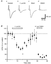
Figure 8. Effects of KA on EPSCs
A, representative EPSCs recorded with the use of the whole-cell patch-clamp technique in the control solution (a), during application of 1 μM KA (b), and after washout (c). These EPSCs were abolished almost completely by further addition of 10 μM CNQX (d). Each trace is the mean of six consecutive EPSCs. The cell was voltage clamped at -71 mV. B, time course of the effect of 1 μM KA on the EPSCs. The relative amplitudes of the EPSCs, with those before KA application as references, are plotted against time (n = 5). KA (1 μM) and CNQX (10 μM) were applied during the period as indicated by the open bars. a, b, c and d in the graph indicate the time points when traces Aa, b, c and d were recorded, respectively.

References

    1. Agrawal S G, Evans R H. The primary afferent depolarizing action of kainate in the rat. British Journal of Pharmacology. 1986;87:345–355. - PMC - PubMed
    1. Baskys A, Malenka R C. Agonists at metabotropic glutamate receptors presynaptically inhibit EPSCs in neonatal rat hippocampus. The Journal of Physiology. 1991;444:687–701. - PMC - PubMed
    1. Bettler B, Mulle C. Neurotransmitter receptors II. AMPA and kainate receptors. Neuropharmacology. 1995;34:123–139. - PubMed
    1. Castillo P E, Malenka R C, Nicoll R A. Kainate receptors mediate a slow postsynaptic current in hippocampal CA3 neurons. Nature. 1997;388:182–186. 10.1038/40645. - DOI - PubMed
    1. Chittajallu R, Vignes M, Dev K K, Barnes J M, Collingridge G L, Henley J M. Regulation of glutamate release by presynaptic kainate receptors in the hippocampus. Nature. 1996;379:78–81. - PubMed

Publication types

MeSH terms

LinkOut - more resources