. 1998 May 26;95(11):5884-90.
doi: 10.1073/pnas.95.11.5884.

Photoactive yellow protein: a structural prototype for the three-dimensional fold of the PAS domain superfamily

Affiliations

Photoactive yellow protein: a structural prototype for the three-dimensional fold of the PAS domain superfamily

J L Pellequer et al. Proc Natl Acad Sci U S A. .

Abstract

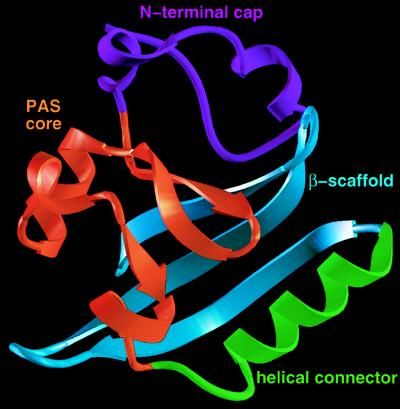
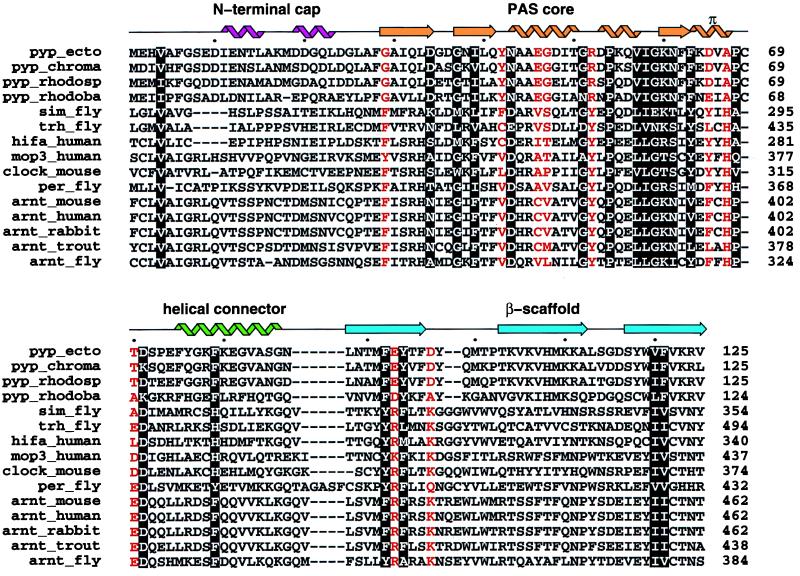
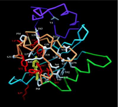
PAS domains are found in diverse proteins throughout all three kingdoms of life, where they apparently function in sensing and signal transduction. Although a wealth of useful sequence and functional information has become recently available, these data have not been integrated into a three-dimensional (3D) framework. The very early evolutionary development and diverse functions of PAS domains have made sequence analysis and modeling of this protein superfamily challenging. Limited sequence similarities between the approximately 50-residue PAS repeats and one region of the bacterial blue-light photosensor photoactive yellow protein (PYP), for which ground-state and light-activated crystallographic structures have been determined to high resolution, originally were identified in sequence searches using consensus sequence probes from PAS-containing proteins. Here, we found that by changing a few residues particular to PYP function, the modified PYP sequence probe also could select PAS protein sequences. By mapping a typical approximately 150-residue PAS domain sequence onto the entire crystallographic structure of PYP, we show that the PAS sequence similarities and differences are consistent with a shared 3D fold (the PAS/PYP module) with obvious potential for a ligand-binding cavity. Thus, PYP appears to prototypically exhibit all the major structural and functional features characteristic of the PAS domain superfamily: the shared PAS/PYP modular domain fold of approximately 125-150 residues, a sensor function often linked to ligand or cofactor (chromophore) binding, and signal transduction capability governed by heterodimeric assembly (to the downstream partner of PYP). This 3D PAS/PYP module provides a structural model to guide experimental testing of hypotheses regarding ligand-binding, dimerization, and signal transduction.


Figures

Figure 1
Figure 1
A proposed PAS/PYP 3D fold illustrated on the PYP structure. The N-terminal cap, colored in purple, contains residues from 1 to 28. The PAS-core, colored in gold, is the domain where higher sequence homology is found among various members of the PAS-containing molecules. It spans from residue 29 to 69. The helical connector, colored in green, includes a short loop followed by the helix α5 and spans residues from 70 to 87. The β-scaffold, colored in blue, contains the last three strands of PYP, spanning residues from 88 to 125.
Figure 2
Figure 2
Similarities revealed by multiple sequence alignment of several members of the PAS-containing proteins and members of the PYP family. The alignment was performed using the program pile-up in the GCG suite (25) starting from the ARNT molecules, then adding each PAS-containing molecule in the list. PYP molecules were manually aligned on the top (see Methods). White letter amino acids are conserved in both PYP and PAS-containing proteins. Red letter amino acids highlight significant differences between PYP and PAS-containing proteins. The secondary structure of PYP is displayed on the top using the color coding from Fig. 1. Helices are represented by “noodles”, strands by arrows and loops by lines. Accession numbers from the SwissProt database, as extracted using the Entrez web service (http://www.ncbi.nlm.nih.gov/Entrez/protein.html) are P16113 (pyp_ecto), X98888 (pyp_rhodosp), X98889 (pyp_rhodoba), M19029 (sim_fly), U33427 (trh_fly), U22431 (hifa_human), U51627 (mop3_human), X03636 (per_fly), U10325 (arnt_mouse), M69238 (arnt_human), D45239 (arnt_rabbit), and AF020426 (arnt_fly). Other sequences were obtained through the Entrez service by searching full names of proteins (pyp_chroma, clock_mouse, and arnt_trout).
Figure 3
Figure 3
Detailed residue interactions for conserved residues that form specific side-chain to main-chain hydrogen bonds in PYP and appear to be retained in PAS-containing molecules. First, Asp 34 OD1 hydrogen bonds to three backbone nitrogens from residues D36, G37, and N38 (2.87 Å, 3.08 Å and 2.89 Å, respectively). Most residues at position 34 have an atom OD1 (Asp, Asn) or similar (OG1 in Ser, Thr). Second, Asn 43 OD1 hydrogen bonds to three backbone nitrogen atoms: A30, A45, and E46 (2.96 Å, 3.55 Å, and 3.06 Å). All residues 43 in Fig. 2 have an atom OD1 or OE1. Third, Asn61 OD1 and ND2 hydrogen bond to three backbone nitrogens and one backbone oxygen: F62, F63, K64, and D36 (3.39 Å, 3.09 Å, 3.00 Å, and 2.96 Å). Almost all residues 61 in Fig. 2 have an atom OD1 or OG1. Drawing made with the program molscript (46).
Figure 4
Figure 4
The PAS-domain of the human ARNT protein modeled from the PYP crystal structure (2phy.pdb). The Cα trace is represented by a tube. The N-terminal cap is colored in magenta, the PAS-core in gold, the helical connector in green, and the β-scaffold in blue. Conserved side chains between PYP and PAS-containing proteins are displayed in white. Amino acids that significantly vary between PYP and PAS-containing molecules are drawn in red. PYP’s chromophore is displayed in yellow in the same orientation as in the PYP molecule. Most conserved residues are located in the hydrophobic core of the PAS-core domain (in gold). Most of significantly variant amino acids (in red) occur in the vicinity of the chromophore pocket as expected from molecules that carry different biological functions. In the ARNT model, H67 and F29 occupy the chromophore pocket. Figure displayed using the Application Visualization System (avs) (AVS, Waltham, MA).
Figure 5
Figure 5
Predicted PAS functional interactions. (A) Amino acid side chains that might participate in a protein–protein interaction are highlighted. The central segment, formed by residues 51-68, is located within 10 Å of the residue Y52 (in yellow). The second area, from residue 95-103 (in cyan), is made of a loop in which an insertion occurs at position 98 in each PAS-containing molecule. The third area is made by two residues (orange) adjacent to the central segment (yellow), H44 and R45, for which their side chains point toward the solvent. (B) same as A but the molecular surface for these residues is displayed.

References

    1. Nambu J R, Lewis J O, Jr, Wharton K A, Crews S T. Cell. 1991;67:1157–1167. - PubMed
    1. Crews S T, Thomas J B, Goodman C S. Cell. 1988;52:143–151. - PubMed
    1. Hoffman E C, Reyes H, Chu F-F, Sander F, Conley L H, Brooks B A, Hankinson O. Science. 1991;252:954–958. - PubMed
    1. Ponting C P, Aravind L. Curr Biol. 1997;7:674–677. - PubMed
    1. Zhulin I B, Taylor B L, Dixon R. Trends Biochem Sci. 1997;22:331–333. - PubMed

Publication types

Substances

LinkOut - more resources