

doi: 10.1073/pnas.95.11.5891.
New methods of structure refinement for macromolecular structure determination by NMR
Affiliations
- PMID: 9600889
- PMCID: PMC34492
- DOI: 10.1073/pnas.95.11.5891
Item in Clipboard
New methods of structure refinement for macromolecular structure determination by NMR
Proc Natl Acad Sci U S A.
.
Abstract
Recent advances in multidimensional NMR methodology have permitted solution structures of proteins in excess of 250 residues to be solved. In this paper, we discuss several methods of structure refinement that promise to increase the accuracy of macromolecular structures determined by NMR. These methods include the use of a conformational database potential and direct refinement against three-bond coupling constants, secondary 13C shifts, 1H shifts, T1/T2 ratios, and residual dipolar couplings. The latter two measurements provide long range restraints that are not accessible by other solution NMR parameters.
Figures
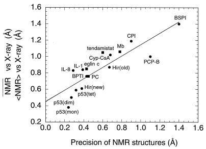
Correlation between backbone precision of NMR
structures and their agreement with x-ray structures. Where the
backbone rms difference between the average NMR coordinates (NMR) and
the corresponding x-ray structures is available, the values are
represented as circles. When only the average backbone rms difference
between an ensemble of NMR structures () and the corresponding
x-ray structure is quoted in the literature, squares are used. The
straight line represents a linear fit to the data with a slope of 0.70,
an intercept of 0.45 Å, and a correlation coefficient of 0.9. The
structures are as follows: p53(mon), p53(dim), and p53(tet) are the
monomer, dimer, and tetramer, respectively, of the p53 oligomerization
domain (51); IL-8, interleukin-8 monomer (52); Hir (new), highly
refined structure of hirudin (53); IL-1, interleukin-1β (6, 7); BPTI,
bovine pancreatic trypsin inhibitor (54); eglin c (55); PC, French bean
plastocyanin (56); tendamistat (57); Hir(old), hirudin (58); Cyp-CsA,
cyclophilin–cyclosporin A complex (59); Mb, carbonmonoxy myoglobin
(helices plus heme; ref. 60); CPI, potato carboxypeptidase inhibitor
(61); PCP-B, procarboxypeptidase B (62); and BSPI, barley serine
proteinase inhibitor 2 (63). The values given exclude conformationally
disordered regions as described in the papers cited. Note that the NMR
structures of IL-8 and Hir(old) were obtained before the corresponding
x-ray structures and that the NMR structure of tendamistat was obtained
independently of and at the same time as the x-ray structure.
Reproduced from ref. .
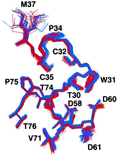
View of the active site and neighboring regions
of reduced human thioredoxin showing a superposition of 40 simulated
annealing structure before (blue) and after (red) 1H
chemical shift refinement. Reproduced from ref. .
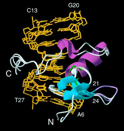
View showing bestfit superpositions of the
restrained regularized mean coordinates obtained with and without
dipolar coupling restraints. The protein is shown as a ribbon diagram
drawn through the Cα positions. The loop between strands
β3 and β4 (residues 21–24) is shown in magenta for the structure
obtained with dipolar coupling restraints and in grey for the structure
obtained without dipolar coupling restraints. Adapted from ref. .
References
-
- Clore G M, Gronenborn A M. Science. 1991;252:1390–1399. - PubMed
-
- Wüthrich K. NMR of Proteins and Nucleic Acids. New York: Wiley; 1986.
-
- Clore G M, Gronenborn A M. Protein Eng. 1987;1:275–288. - PubMed
-
- Dyson H J, Gippert G P, Case D A, Holmgren A, Wright P E. Biochemistry. 1990;29:4129–4136. - PubMed
-
- Forman-Kay J D, Clore G M, Wingfield P T, Gronenborn A M. Biochemistry. 1991;30:2685–2698. - PubMed
MeSH terms
Substances
LinkOut - more resources
Full Text Sources
