

doi: 10.1073/pnas.95.11.5935.
Architecture and mechanism of the light-harvesting apparatus of purple bacteria
Affiliations
- PMID: 9600895
- PMCID: PMC34498
- DOI: 10.1073/pnas.95.11.5935
Item in Clipboard
Architecture and mechanism of the light-harvesting apparatus of purple bacteria
Proc Natl Acad Sci U S A.
.
Abstract
Photosynthetic organisms fuel their metabolism with light energy and have developed for this purpose an efficient apparatus for harvesting sunlight. The atomic structure of the apparatus, as it evolved in purple bacteria, has been constructed through a combination of x-ray crystallography, electron microscopy, and modeling. The detailed structure and overall architecture reveals a hierarchical aggregate of pigments that utilizes, as shown through femtosecond spectroscopy and quantum physics, elegant and efficient mechanisms for primary light absorption and transfer of electronic excitation toward the photosynthetic reaction center.
Figures
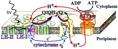
Schematic representation of the photosynthetic
apparatus in the intracytoplasmic membrane of purple bacteria. The RC
(red) is surrounded by the light-harvesting complex I (LH-I, green) to
form the LH-I–RC complex, which is surrounded by multiple
light-harvesting complexes LH-II (green), forming altogether the PSU.
Photons are absorbed by the light-harvesting complexes and excitation
is transferred to the RC initiating a charge (electron-hole)
separation. The RC binds quinone QB, reduces it to hydroquinone
QBH2, and releases the latter.
QBH2 is oxidized by the bc1 complex,
which uses the exothermic reaction to pump protons across the membrane;
electrons are shuttled back to the RC by the cytochrome
c2 complex (blue) from the
ubiquinone–cytochrome bc1 complex (yellow). The electron
transfer across the membrane produces a large proton gradient that
drives the synthesis of ATP from ADP by the ATPase (orange). Electron
flow is represented in blue, proton flow in red, and quinone flow,
likely confined to the intramembrane space, in black.
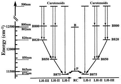
Energy levels of the electronic excitations in
the PSU of BChl a containing purple bacteria. The diagram
illustrates a funneling of excitation energy toward the photosynthetic
RC. The dashed lines indicate (vertical) intracomplex excitation
transfer, and the solid lines (diagonal) indicate intercomplex
excitation transfer. LH-I exists in all purple bacteria; LH-II exists
in most species; LH-III arises in certain species only.
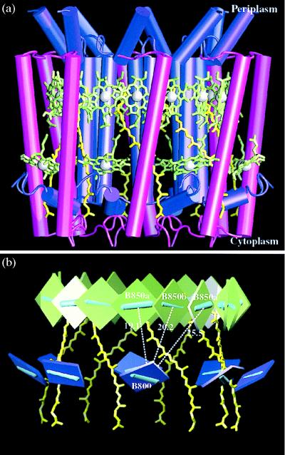
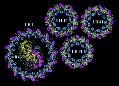
The octameric LH-II complex from Rs.
molischianum (19). (a) The α-helical segments
are represented as cylinders with the α-apoproteins (inside) in blue
and the β-apoprotein (outside) in magenta. The BChl molecules are in
green with phytyl tails truncated for clarity. The lycopenes are in
yellow. (b) Arrangement of chromophores with BChls
represented as squares, and with carotenoids (lycopenes) in a licorice
representation. Bars connected with the BChls represent the Qy
transition dipole moments as defined by the vector connecting the N
atom of pyrrol I and the N atom of pyrrol III (22). Representative
distances between central Mg atoms of B800 BChl and B850 BChl are given
in Å. The B850 BChls bound to the α-apoprotein and the
β-apoprotein are denoted as B850a and B850b, respectively; BChl
B850a′ is bound to the (left) neighboring heterodimer.
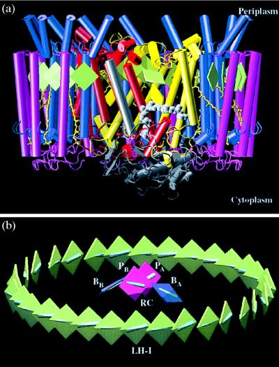
Structure of the LH-I–RC complex.
(a) Side view of the LH-I–RC complex with three LH-I
αβ-heterodimers on the front side removed to expose the RC in the
interior. The α-helices are represented as cylinders with the L, M,
and H subunits of the RC in yellow, red, and gray, and the
α-apoprotein and the β-apoprotein of the LH-I in blue and magenta.
BChls and bacteriopheophytins are represented as green and yellow
squares, respectively. Carotenoids (spheroidenes) are in a yellow
licorice representation, and quinone QB is rendered by gray van
der Waals spheres. QB shuttles in and out (as
QBH2) of the LH-I–RC complex as indicated in Fig.
1. (b) Arrangement of BChls in the LH-I–RC complex. The
BChls are represented as squares with B875 BChls of LH-I in green, and
the special pair (PA and PB) and the accessory BChls
(BA and BB) of the RC in red and blue, respectively;
cyan bars represent Qy transition moments of BChls. [Produced
with the program vmd (25)].
Arrangement of pigment–protein complexes in the
modeled bacterial PSU of Rb. sphaeroides. The
α-helices are represented as Cα-tracing tubes with
α-apoproteins of both LH-I and LH-II in blue and β-apoproteins in
magenta, and the L, M, and H subunits of RC in yellow, red, and gray,
respectively. All the BChls are in green, and carotenoids are in
yellow. [Produced with the program vmd (25)].
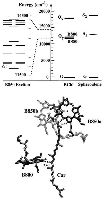
BChl–carotenoid interactions. (Upper
Left) Exciton bands of the circular B850 BChl aggregate as
determined by quantum chemical (INDO/S) calculations (29) based on
coordinates of the crystal structure of LH-II from Rs.
molischianum (19). The degenerate states that carry all the
oscillator strength are highlighted by thickened lines. (Upper
Right) Excitation energies of BChl and carotenoid states in
LH-II of Rb. sphaeroides. Solid lines represent
spectroscopically measured energy levels. The dashed line indicates the
estimated (see refs. and 47) energy for the optically forbidden
S1 state of the carotenoid spheroidene.
(Lower) Arrangement of spheroidene and the most
proximate BChls based on the modeled structure of LH-II from Rb.
sphaeroides. Close contacts between BChl and the carotenoid
spheroidene are indicated by representative distances (in angstroms).
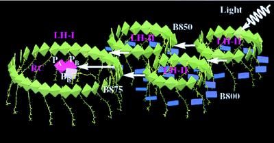
Excitation transfer in the bacterial
photosynthetic unit. LH-II contains two types of BChls, commonly
referred to as B800 (dark blue) and B850 (green), which absorb at 800
nm and 850 nm, respectively. BChls in LH-I absorb at 875 nm and are
labeled B875 (green). PA and PB refer to the RC special
pair, and BA, BB refer to the accessory BChls in the
RC. The figure demonstrates the coplanar arrangement of the B850 BChl
ring in LH-II, the B875 BChl ring of LH-I, and the RC BChls PA,
PB, BA, BB. [Produced with the program
vmd (25)].
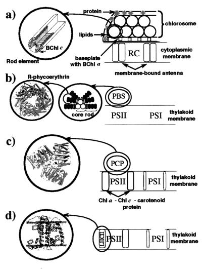
Schematic representation of proposed models of
the PSUs in other photosynthetic systems. The figure displays inter-
and extramembrane light-harvesting complexes, together with the RCs (RC
in green bacteria, and PS-I and PS-II in cyanobacteria,
dinoflagellates, and green plants). (a) Green bacteria:
The major light-harvesting complex, chlorosome, contains rod-like BChl
c aggregates surrounded by a layer of protein embedding
lipids. Excitation energy harvested by the rod-like aggregates reaches
the RC through a BChls a containing baseplate and
membrane-bound light-harvesting BChl a complexes.
(b) Cyanobacteria: The dominant light-harvesting complex
of cyanobacteria and red algae, phycobilisome (PBS), is unique in
choosing linear tetrapyrroles as pigments. Several types of disk-like
pigment–protein complexes such as R-phycoerythrin (51) constitute the
phycobilisome rods and core. (c) Dinoflagellates: The
photosynthetic unit of dinoflagellates consists of several
membrane-bound pigment–protein complexes and an extramembrane
light-harvesting complex, the peridinin–chlorophyll–protein (PCP).
(d) Green plants: Chloroplasts of green plants possess
chlorophyll-carotenoid containing LHCII (6) as the most abundant
light-harvesting complex. [Images of R-phycoerythrin and PCP were
produced with the program vmd (25)].
References
-
- Hu X, Schulten K. Physics Today. 1997;50:28–34.
-
- Duysens L N M. Ph.D. thesis. The Netherlands: Utrecht; 1952.
-
- Cogdell R, Fyfe P, Barrett S, Prince S, Freer A, Isaacs N, McGlynn P, Hunter C. Photosynth Res. 1996;48:55–63. - PubMed
-
- Krauss N, Schubert W-D, Klukas O, Fromme P, Witt H T, Saenger W. Nat Struct Biol. 1996;3:965–973. - PubMed
Publication types
MeSH terms
Substances
Grants and funding
LinkOut - more resources
Full Text Sources
