. 1998 May 26;95(11):6055-60.
doi: 10.1073/pnas.95.11.6055.

Nonneuronal isoforms of STOP protein are responsible for microtubule cold stability in mammalian fibroblasts

Affiliations

Nonneuronal isoforms of STOP protein are responsible for microtubule cold stability in mammalian fibroblasts

E Denarier et al. Proc Natl Acad Sci U S A. .

Abstract

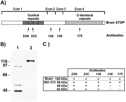
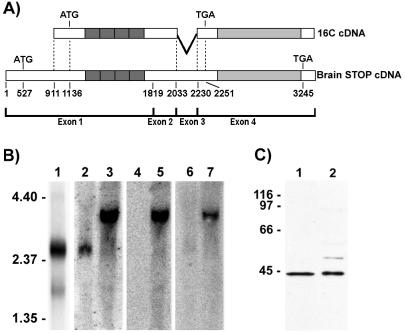
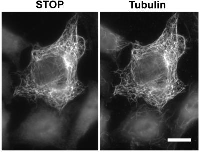
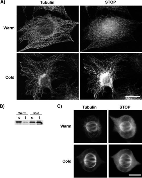
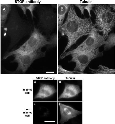
A number of cycling mammalian cells, such as NIH 3T3, contain abundant subsets of cold-stable microtubules. The origin of such microtubule stabilization in nonneuronal cells is unknown. We have previously described a neuronal protein, stable tubule-only polypeptide (STOP), that binds to microtubules and induces cold stability. We find that NIH 3T3 fibroblasts contain a major 42-kDa isoform of STOP (fibroblastic STOP, F-STOP). F-STOP contains the central repeats characteristic of brain STOP but shows extensive deletions of N- and C-terminal protein domains that are present in brain STOP. These deletions arise from differences in STOP RNA splicing. Despite such deletions, F-STOP has full microtubule stabilizing activity. F-STOP accumulates on cold-stable microtubules of interphase arrays and is present on stable microtubules within the mitotic spindle of NIH 3T3 cells. STOP inhibition by microinjection of affinity-purified STOP central repeat antibodies into NIH 3T3 cells abolishes both interphase and spindle microtubule cold stability. Similar results were obtained with Rat2 cells. These results show that STOP proteins have nonneuronal isoforms that are responsible for the microtubule cold stability observed in mammalian fibroblasts.


Figures

Figure 1
Figure 1
STOP antigens in NIH 3T3 cell extracts. (A) Schematic representation of mouse brain STOP showing domain structure of the protein. Protein domains encoded by the four exons are indicated. Mouse brain STOP contains a central repeat domain (encoded by exon 1) composed of four repeats and a C-terminal repeat domain (encoded by exon 4) composed of 28 repeats. The localization of the epitopes recognized by STOP antibodies 23N, 23C, 136, 139, and 175 is indicated. (B) Immunoblot analysis of NIH 3T3 cell extract (lane 1) and mouse brain extract (lane 2) using 23C STOP antibody. NIH 3T3 cell extract (40 μg) and brain extract (4 μg) were separated by SDS/PAGE on 8% gels. Size markers are in kDa. (C) Reactivity of brain and NIH 3T3 cell STOP proteins with the various STOP antibodies. For each STOP species, presence (+) or absence (−) of reactivity with the different STOP antibodies is indicated.
Figure 2
Figure 2
Characterization of the F-STOP cDNA. (A) Schematic representation showing alignment of 16C cDNA with mouse brain STOP cDNA. Nucleotides are numbered according to the mouse brain STOP cDNA sequence, as deduced from the mouse STOP gene. 16C cDNA lacks nucleotides 1–910 of the brain STOP cDNA and also lacks the entire sequence of exon 3. (B) Northern blot analysis of RNAs from NIH 3T3 cells and from mouse brain. Poly(A)+ RNAs (3 μg) from NIH 3T3 cells were hybridized with 32P-labeled 16C cDNA, and a major 2.4-kb RNA species (lane 1) was detected. Total NIH 3T3 cell RNAs (30 μg; lanes 2, 4, and 6) or total mouse brain RNAs (30 μg; lanes 3, 5, and 7) were hybridized with 32P-labeled probes corresponding to the 3′ region of exon 1 (positions 787–1,818; lanes 2 and 3), to the 5′ region of exon 1 (positions 77–787; lanes 4 and 5), and to exon 3 (positions 2,044–2,181; lanes 6 and 7). As with 16C cDNA, the major STOP mRNA in NIH 3T3 cells lacked nucleotide sequences corresponding to the 5′ end of the brain STOP cDNA and the entire sequence of exon 3. Size markers are in kilobases. (C) Comparison of the translation product of p16C-Apa with F-STOP. Reticulocyte lysate reactions (5 μl; lane 1) and NIH 3T3 cell extracts (30 μg; lane 2) were separated by SDS/PAGE on 10% gels and immunoblotted with 23C STOP antibody. The translation product of p16C-Apa comigrated with F-STOP. Size markers are in kDa.
Figure 3
Figure 3
F-STOP induces microtubule cold stability in HeLa cells. HeLa cells were transfected with cDNA coding for F-STOP. After 24 h, cells were exposed to cold temperature, lysed, and then double-stained with STOP antibody 23C (Left) and with tubulin antibody TUB 2.1 (Right). F-STOP associates with microtubules in transfected cells and F-STOP-associated polymers are resistant to depolymerization induced by cold. (Bar = 10 μm.)
Figure 4
Figure 4
STOP localization in NIH 3T3 cells. (A) NIH 3T3 cells either were immediately fixed (row warm) or were first exposed to cold temperature for 30 min (row cold). Cells were then double-stained using mAb TUB 2.1 to label tubulin (Left) and 23C antibody to label STOP proteins (Right). STOP staining of microtubules is strongly enhanced in cold-treated cells as compared with untreated cells. (Bar = 10 μm.) (B) Immunoblot analysis of F-STOP presence in Triton-soluble cell fraction (lanes s) and Triton-insoluble cell fraction (lanes i). NIH 3T3 cells were immediately extracted with a Triton-based buffer (lanes warm) or first incubated at 0°C for 30 min (lanes cold). Cell extracts were then separated by SDS/PAGE on 8% gels and assayed for F-STOP content on immunoblots using 23C STOP antibody. In repeated experiments, patterns of F-STOP distribution among soluble and insoluble fractions from the same cells were highly reproducible. The apparent difference in total STOP content between cold-treated and untreated cells reflects random variations between preparations. (C) Mitotic cells analyzed in the same manner as described in A. (Bar = 5 μm.)
Figure 5
Figure 5
Interphase and spindle microtubule disassembly in cold-treated cells injected with STOP antibody. NIH 3T3 cells were injected with affinity-purified 23C STOP antibody. After a 2-h recovery period, cells were exposed to cold temperature for 30 min. Cells were then fixed and double-stained with Cy3-conjugated anti-rabbit antibody to detect injected 23C antibody (A, C, and E) and mAb TUB 2.1-fluorescein isothiocyanate anti-mouse antibody to detect tubulin (B, D, and F). (A and B) Injected and noninjected interphase cells. (C and D) Injected metaphase cell. (E and F) Noninjected metaphase cell. STOP inhibition abolishes microtubule cold stability. (Bars = 10 μm.)

References

    1. McNally F J. Curr Opin Cell Biol. 1996;8:23–29. - PubMed
    1. Salmon E D, Leslie R J, Saxton W M, Karow M L, McIntosh J R. J Cell Biol. 1984;99:2165–2174. - PMC - PubMed
    1. Saxton W M, Stemple D L, Leslie R J, Salmon E D, Zavortink M, McIntosh J R. J Cell Biol. 1984;99:2175–2186. - PMC - PubMed
    1. Belmont L D, Hyman A A, Sawin K E, Mitchison T J. Cell. 1990;62:579–589. - PubMed
    1. Mitchison T J, Kirschner M. Nature (London) 1984;312:237–242. - PubMed

Publication types

Associated data