

doi: 10.1073/pnas.95.11.6118.
Huntingtin aggregation monitored by dynamic light scattering
Affiliations
- PMID: 9600927
- PMCID: PMC27595
- DOI: 10.1073/pnas.95.11.6118
Item in Clipboard
Huntingtin aggregation monitored by dynamic light scattering
Proc Natl Acad Sci U S A.
.
Abstract
An initial stage of fibrillogenesis in solutions of glutathione S-transferase-huntingtin (GST-HD) fusion proteins has been studied by using dynamic light scattering. Two GST-HD systems with poly-L-glutamine (polyGln) extensions of different lengths (20 and 51 residues) have been examined. For both systems, kinetics of z-average translation diffusion coefficients (Dapp) and their angular dependence have been obtained. Our data reveal that aggregation does occur in both GST-HD51 and GST-HD20 solutions, but that it is much more pronounced in the former. Thus, our approach provides a powerful tool for the quantitative assay of GST-HD fibrillogenesis in vitro.
Figures
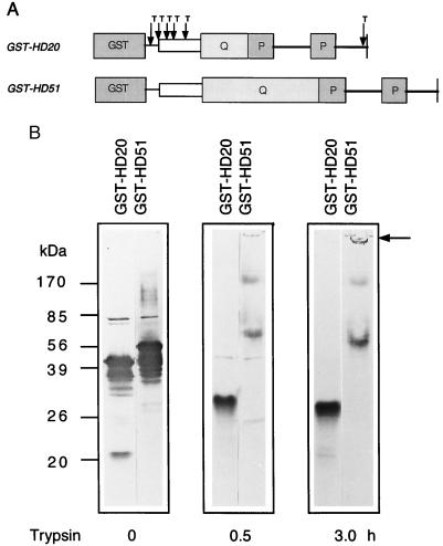
(A) Schematic representation of primary structure of GST-HD fusion proteins. The amino acid sequence corresponding to exon 1 of huntingtin is boxed. Q and P stand for polyglutamine and polyproline extensions, respectively. Arrows and T indicate cleavage sites for trypsin. (B) Site-specific proteolysis of GST-HD fusion proteins with trypsin. Tryptic digestions were performed at 37°C for the indicated times. Undigested and trypsin-digested proteins were subjected to 12.5% SDS/PAGE, blotted onto nitrocellulose membranes, and probed with the anti-huntingtin antibody HD1. Arrow marks the origin of electrophoresis.
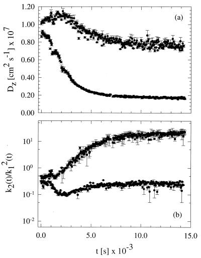
(a) Apparent diffusion coefficient Dapp(t) plotted as function of time for selected fusion protein preparations GST-HD20 (•) and GST-HD51 (▪). Measurements were conducted at a fixed scattering angle, 45°. (b) Normalized second cumulant of GST-HD20 (•) and GST-HD51 (▪) as a function of time. Note that the 5-fold drop of the apparent diffusion coefficient is accompanied by a 100-fold increment of the normalized second cumulant, which can be understood as a drastic increment of the solution polydispersity.
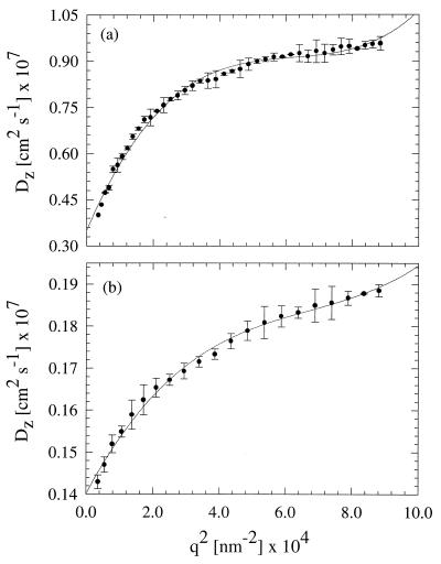
Angular dependence of the z-average diffusion coefficient of the (a) GST-HD20 and (b) GST-HD51 protein preparations. SDs were estimated from an average of three experiments conducted 18 hr after collecting the time-resolved results shown in Fig. 2. Note that the q2 dependence of Dapp for both samples deviates appreciably from linearity. Further, the GST-HD20 sample exhibits roughly a two times higher limiting Dapp(q) value than the GST-HD51 one.
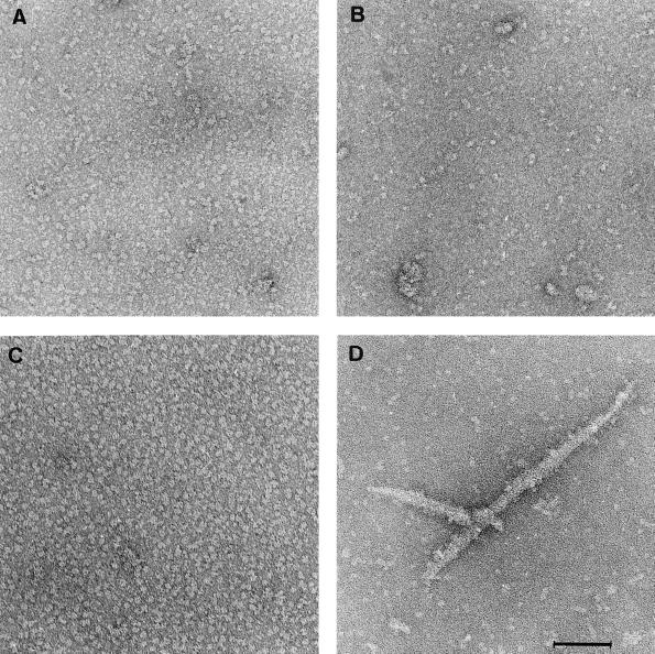
Electron micrographs of GST-HD20 (A and B) and GST-HD51 (C and D) proteins before (A and C) and after (B and D) treatment with trypsin. Samples were negatively stained with 1% uranyl acetate. (Scale bar = 100 nm.)
References
-
- Harper P S, editor. Huntington Disease. 22nd Ed. London: Saunders; 1991.
-
- The Huntington Disease Collaborative Research Group. Cell. 1993;72:971–983. - PubMed
-
- Sathasivam K, Amaechi I, Mangiarini L, Bates G P. Hum Genet. 1997;99:692–695. - PubMed
-
- Trottier Y, Lutz Y, Stevanin G, Imbert G, Devys D, Cancel G, Sandou F, Weber C, David G, Tora L, et al. Nature (London) 1995;378:403–406. - PubMed
Publication types
MeSH terms
Substances
LinkOut - more resources
Full Text Sources
Other Literature Sources
Medical
Research Materials
