. 1998 May 26;95(11):6134-8.
doi: 10.1073/pnas.95.11.6134.

Visualization of elongation factor G on the Escherichia coli 70S ribosome: the mechanism of translocation

Affiliations

Visualization of elongation factor G on the Escherichia coli 70S ribosome: the mechanism of translocation

R K Agrawal et al. Proc Natl Acad Sci U S A. .

Abstract

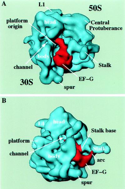
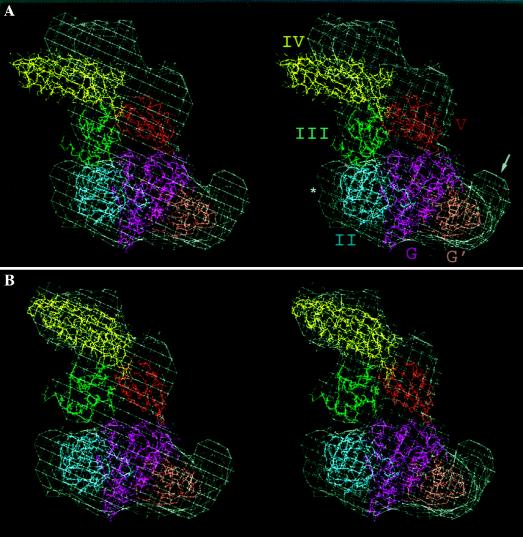
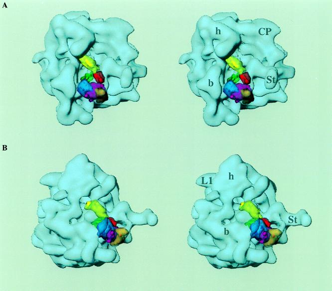
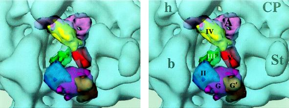
During protein synthesis, elongation factor G (EF-G) binds to the ribosome and promotes the step of translocation, a process in which tRNA moves from the A to the P site of the ribosome and the mRNA is advanced by one codon. By using three-dimensional cryo-electron microscopy, we have visualized EF-G in a ribosome-EF-G-GDP-fusidic acid complex. Fitting the crystal structure of EF-G-GDP into the cryo density map reveals a large conformational change mainly associated with domain IV, the domain that mimics the shape of the anticodon arm of the tRNA in the structurally homologous ternary complex of Phe-tRNAPhe, EF-Tu, and a GTP analog. The tip portion of this domain is found in a position that overlaps the anticodon arm of the A-site tRNA, whose position in the ribosome is known from a study of the pretranslocational complex, implying that EF-G displaces the A-site tRNA to the P site by physical interaction with the anticodon arm.


Figures

Figure 1
Figure 1
The 3D cryo electron microscope maps of E. coli 70S ribosome complexed with EF-G–GDP–fusidic acid. (A) Side view showing the binding position of EF-G (red) in the intersubunit space. The 30S subunit is on the left and 50S subunit is on the right. EF-G is directly visible and seen to interact with both 30S and 50S subunits (6, 7). The tip of the elongated portion of EF-G is situated in the neck region of 30S subunit, where the anticodon loop region of A site tRNA binds (11, 12). (B) The 30S side view, showing the arc-like structure formed between the EF-G and a protusion at the base of the L7/L12 stalk. Note the clearly defined well-extended L7/L12 stalk. In this view, the tip (domain IV) of EF-G can be seen through the 30S channel (18) where the anticodon of A site tRNA in a pretranslocational complex binds (ref ; R.K.A., P.P., R.A.G., N. Burkhardt, R. Jünemann, K. H. Nierhaus, and J.F., unpublished results; also see Fig. 4). Landmarks: L1, protein L1 of the 50S subunit; arc, arc-like connection between EF-G and an extension from the stalk base (see text).
Figure 2
Figure 2
Stereo view showing the fitting of the crystal structure (9) of EF-G–GDP into the extra mass of density corresponding to EF-G. The density map corresponding to EF-G is shown in white wire-mesh form. The various domains of the crystal structure are shown in different colors. (A) Fitting using domains I and II as the main guide to align the whole crystal structure into the density map (see text). (B) Separate fitting of the substructure formed by domains III–V (see text). To obtain an optimum fit, this structure was shifted by approximately 10 Å toward the central protuberance of the 50S subunit and rotated by about 10°, around a pivoting point in the contact region between domains II and III, toward the L1 side, whereas domains I and II were left in the same position as in A. The arrow points to the portion of the density map that is partially occupied by the G′ domain and involved in making the arc-like connection with the L7/L12 stalk (see also Figs. 1B and 3B). The asterisk shows a partially unoccupied region that probably comes from the ribosome because of a conformational change at the site of interaction with domain II.
Figure 3
Figure 3
Stereo view of a low-resolution version of the EF-G x-ray structure shown in Fig. 2B, superimposed onto the 3D map of the 70S–EF-G–GDP–fusidic acid complex (shown as a transparent blue surface). Color coding for different domains of the x-ray structure is the same as in Fig. 2: domain I, magenta and brown (core G domain, magenta; insert G′ domain, brown); domain II, blue; domain III, green; domain IV, yellow; and domain V, red. (A) In the intersubunit face view, the 30S subunit is on the left and the 50S subunit on the right. (B) Same as a but presented from the 30S-solvent side to show that the G′ domain is involved in the formation of the arc-like structure with the base of the L7/L12 stalk. Because only the lower part of the arc-like structure is occupied by the x-ray structure, the upper part is probably contributed by the C-terminal domain of one of the folded L7/L12 molecule (see text), which becomes pronounced only upon factor binding (see also ref. 24). Landmarks have been abbreviated to avoid visual complexity of the stereo picture. For 30S subunit: h, head; b, body. For 50S subunit: CP, central protuberance; St, L7/L12 Stalk; L1, L1 protein.
Figure 4
Figure 4
Same as Fig. 3A but showing a magnified view of the site of interaction of different domains of EF-G with the ribosome. Particularly important to note is the partial overlap of domain IV with the A-site tRNA (pink), as obtained from a cryo map of a pretranslocational complex (R.K.A., P.P., R.A.G., N. Burkhardt, R. Jünemann, K. H. Nierhaus, and J.F., unpublished results). Also, note the proximity of the anticodon end of the A site tRNA to the tip of the domain IV. Various domains are labeled with roman numerals and the A-site tRNA is marked as A. Other landmarks are as in Fig. 3.

Comment in

Similar articles

Cited by

References

    1. Nishizuka Y, Lipmann F. Proc Natl Acad Sci USA. 1966;55:212–219. - PMC - PubMed
    1. Kawakita M, Arai K, Kaziro Y. J Biochem. 1974;76:801–809. - PubMed
    1. Chinali G, Parmeggiani A. Eur J Biochem. 1982;125:415–421. - PubMed
    1. Voigt J, Nagel K. J Biol Chem. 1993;268:100–106. - PubMed
    1. Rodnina M V, Savelsbergh A, Katunin V I, Wintermeyer W. Nature (London) 1997;385:37–41. - PubMed

Publication types

MeSH terms

Substances

LinkOut - more resources