. 1998 May 26;95(11):6169-74.
doi: 10.1073/pnas.95.11.6169.

Tyrosine kinase-dependent activation of a chloride channel in CD95-induced apoptosis in T lymphocytes

Affiliations

Tyrosine kinase-dependent activation of a chloride channel in CD95-induced apoptosis in T lymphocytes

I Szabò et al. Proc Natl Acad Sci U S A. .

Abstract

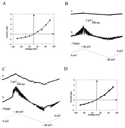
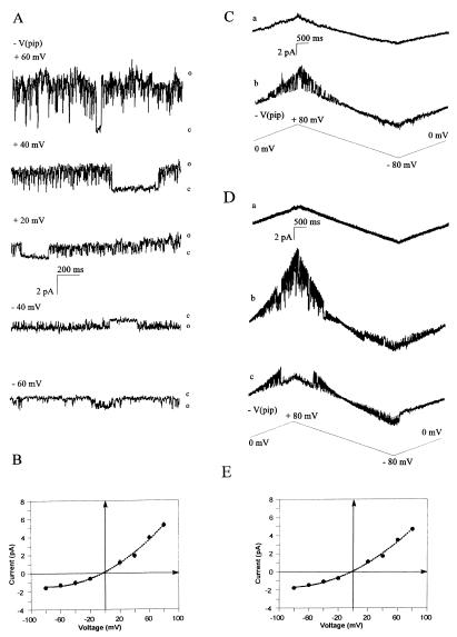
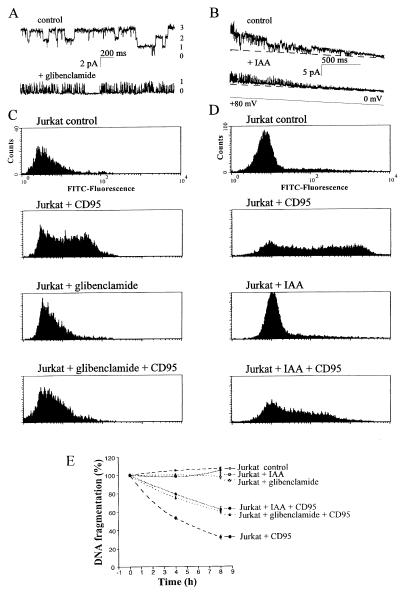
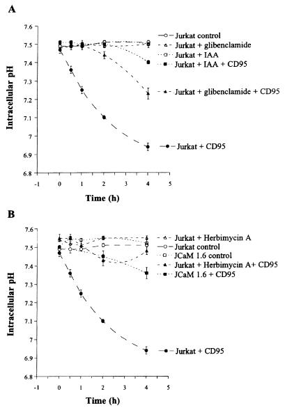
CD95/Fas/APO-1 mediated apoptosis is an important mechanism in the regulation of the immune response. Here, we show that CD95 receptor triggering activates an outwardly rectifying chloride channel (ORCC) in Jurkat T lymphocytes. Ceramide, a lipid metabolite synthesized upon CD95 receptor triggering, also induces activation of ORCC in cell-attached patch clamp experiments. Activation is mediated by Src-like tyrosine kinases, because it is abolished by the tyrosine kinase inhibitor herbimycin A or by genetic deficiency of p56lck. In vitro incubation of excised patches with purified p56lck results in activation of ORCC, which is partially reversed upon addition of anti-phosphotyrosine antibody. Inhibition of ORCC by four different drugs correlates with a 30-65% inhibition of apoptosis. Intracellular acidification observed upon CD95 triggering is abolished by inhibition of either ORCC or p56lck. The results suggest that tyrosine kinase-mediated activation of ORCC may play a role in CD95-induced cell death in T lymphocytes.


Figures

Figure 1
Figure 1
ORCC is activated by CD95 ligation. (A) Open channel I-V relation of ORCC activity in inside-out excised patches in control condition. ORCC was activated by patch excision and depolarization. Error bars represent SEM based on n ≥ 20 amplitudes measured per potential from 10 different experiments. For every current trace recorded in excised patches voltages are expressed with reference to the pipette solution. (B) ORCC activity in cell-attached configuration before (trace a) and after perfusion of the cells with 200 ng/ml anti-human CD95 antibody for 11 min (trace b). Voltage ramps were applied as indicated at 60-sec intervals. For every current trace recorded in on-cell patches voltages are expressed relative to the resting potential. Between the ramps patches were held at resting potential for all cell-attached recordings and at 0 mV for all excised patches. (C) Anti-mouse CD95 antibody does not stimulate ORCC in human Jurkat T lymphocytes in the cell-attached patch configuration after 20 min of incubation (trace a). After excision and depolarization ORCC is activated (trace b). (D) Open channel I-V curve of channels recorded in cell-attached configuration after stimulation of the cells with 200 ng/ml anti-human CD95 antibody (▪) or with 200 μM dibutyryl-cAMP (•). All data are expressed as arithmetic mean ± SEM (n ≥ 14 for each point, obtained from nine and seven experiments, respectively).
Figure 2
Figure 2
ORCC is activated by a signaling cascade involving ceramides and Src-like tyrosine kinases. (A) Perfusion with C6-ceramide (20 μM) results in activation of ORCC. Shown are representative current traces at steady-state potentials in cell-attached configuration 7 min after C6 ceramide addition. In the experiment shown the open probability of the channel increased from 0 to 0.93 ± 0.4 at 8 different voltages. Traces were selected to show channel closures. The activity upon ceramide treatment did not show significant voltage dependence in most experiments. Open and closed levels are indicated. (B) I-V curves of channels activated by 20 M C6-ceramide in cell-attached configuration. All data are expressed as arithmetic mean ± SEM (n ≥ 18 for each point, obtained from nine different experiments). (C) Pretreatment of Jurkat with 10 μM herbimycin A for 10 h prevents ORCC activation upon perfusion with 200 ng/ml anti-human CD95 antibody. Shown is a current trace in cell-attached configuration (trace a) from a herbimycin A-pretreated cell incubated with anti-CD95 for 17 min and a trace (b) from the same patch after patch excision and depolarization demonstrating the presence of ORCC. (D) Addition of purified p56lck to excised patches activates ORCC (trace b). Active ORCC is inactivated by the addition of anti-phosphotyrosine antibodies 4G10. Shown is a representative current trace from an inside-out excised patch held at 0 mV before (trace a) and 5 min after the addition of 2 units purified p56lck + 10 μM ATP (trace b). Trace c was recorded from the same patch after perfusion of the patch chamber with 10 μg/ml antiphosphotyrosine antibodies 4G10 (in the presence of p56lck and ATP). Upon addition of p56lck two ORCC were active (b) whereas only one channel remained relatively active after the addition of 4G10 (c) in the same patch. (E) Open channel current–voltage relation of ORCC activated by purified p56lck in inside-out excised patches. Error bars represent SEM based on n ≥ 12 amplitudes measured per potential from six different experiments.
Figure 3
Figure 3
Glibenclamide and IAA block ORCC and CD95 induced apoptosis. (A) Displayed are traces from an inside-out excised patch held at +40 mV in control condition (Upper) and 2 min after perfusion of the chamber with 100 μM glibenclamide (Lower). The results are representative for three experiments. (B) Representative current traces from an inside-out excised patch before (Upper) and after 8-min (Lower) incubation with 100 μM IAA. Dashed line shows closed level. (C) Treatment of Jurkat cells with 100 μM glibenclamide inhibits CD95-triggered apoptosis determined by binding of FITC-Annexin V and fluorescence-activated cell sorter analysis. Cells were incubated for 4 h with 200 ng/ml anti-human CD95 antibody in the presence or absence of glibenclamide. Shown are representative histograms of FITC-fluorescence. Apoptosis was reduced by 63 ± 4% in six independent experiments. (D) IAA (100 μM) reduces CD95-triggered apoptosis. Apoptosis was determined as in C. Shown are representative histograms of four experiments. (E) Treatment of Jurkat cells with 100 μM glibenclamide or 100 μM IAA reduces CD95-triggered apoptosis, measured by fragmentation of [3H]thymidine-labeled DNA. All data are expressed as arithmetic mean ± SEM (n = 9 for each point). Differences between CD95-triggered apoptosis in the absence and in the presence of inhibitors are significant at 4 and 8 h of incubation (P < 0.05, two-tailed t test).
Figure 4
Figure 4
Cellular acidification upon CD95 stimulation is prevented by glibenclamide and IAA (A) or by genetic deficiency or pharmacological inhibition of Src-like tyrosine kinases (B). (A) Treatment of Jurkat cells with 200 ng/ml anti-human CD95 antibody induces a time-dependent reduction of intracellular pH. Cellular acidification is inhibited by 100 μM glibenclamide or by 100 μM IAA. All data are expressed as arithmetic mean ± SEM (n = 20–60 for each point). (B) Genetic deficiency of p56lck in JCaM1.6 cells or inhibition of Src-like tyrosine kinases by preincubation with 10 μM herbimycin A for 10 h prevents cellular acidification upon stimulation with 200 ng/ml anti-human CD95. Differences are significant (P < 0.05, two-tailed t test) 30 min after CD95 triggering. All data are expressed as arithmetic mean ± SEM (n = 20–60 for each point).

Similar articles

Cited by

References

    1. Thompson C B. Science. 1995;267:1456–1462. - PubMed
    1. Nagata S, Goldstein P. Science. 1995;267:1449–1456. - PubMed
    1. Singer G G, Abbas A K. Immunity. 1994;1:365–372. - PubMed
    1. Gulbins E, Szabò I, Lang F. Cell Physiol Biochem. 1996;6:361–375.
    1. Nagata S. Cell. 1997;88:355–365. - PubMed

Publication types