

Silicatein alpha: cathepsin L-like protein in sponge biosilica
- PMID: 9600948
- PMCID: PMC27641
- DOI: 10.1073/pnas.95.11.6234
Silicatein alpha: cathepsin L-like protein in sponge biosilica
Abstract
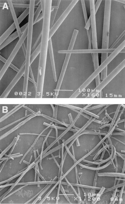
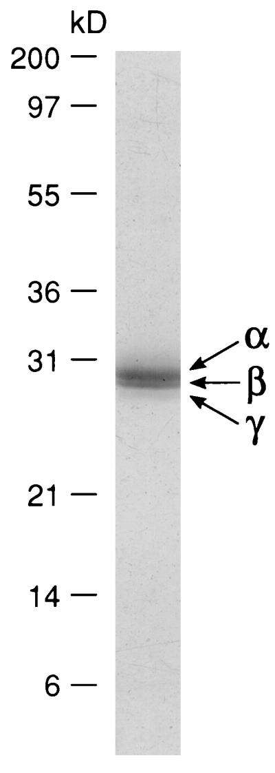
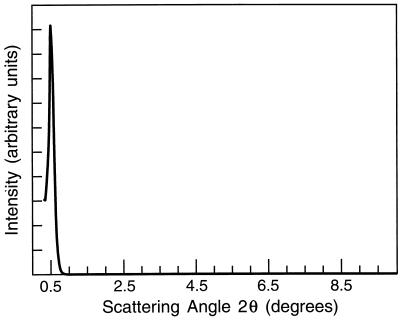
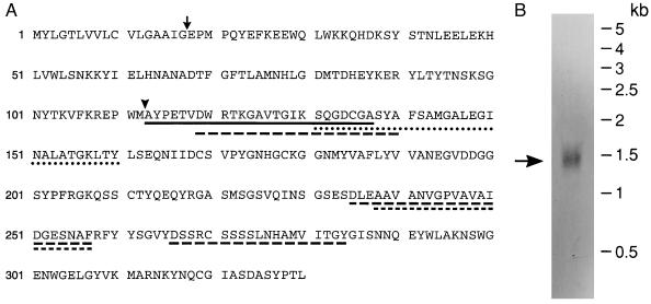
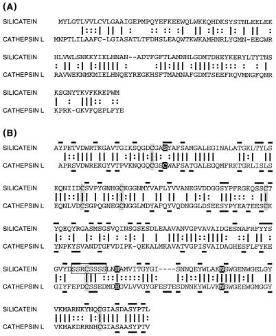
Earth's biota produces vast quantities of polymerized silica at ambient temperatures and pressures by mechanisms that are not understood. Silica spicules constitute 75% of the dry weight of the sponge Tethya aurantia, making this organism uniquely tractable for analyses of the proteins intimately associated with the biosilica. Each spicule contains a central protein filament, shown by x-ray diffraction to exhibit a highly regular, repeating structure. The protein filaments can be dissociated to yield three similar subunits, named silicatein alpha, beta, and gamma. The molecular weights and amino acid compositions of the three silicateins are similar, suggesting that they are members of a single protein family. The cDNA sequence of silicatein alpha, the most abundant of these subunits, reveals that this protein is highly similar to members of the cathepsin L and papain family of proteases. The cysteine at the active site in the proteases is replaced by serine in silicatein alpha, although the six cysteines that form disulfide bridges in the proteases are conserved. Silicatein alpha also contains unique tandem arrays of multiple hydroxyls. These structural features may help explain the mechanism of biosilicification and the recently discovered activity of the silicateins in promoting the condensation of silica and organically modified siloxane polymers (silicones) from the corresponding silicon alkoxides. They suggest the possibility of a dynamic role of the silicateins in silicification of the sponge spicule and offer the prospect of a new synthetic route to silica and siloxane polymers at low temperature and pressure and neutral pH.
Figures
Similar articles
-
Silicateins, the major biosilica forming enzymes present in demosponges: protein analysis and phylogenetic relationship.Gene. 2007 Jun 15;395(1-2):62-71. doi: 10.1016/j.gene.2007.02.014. Epub 2007 Feb 28. Gene. 2007. PMID: 17408887
-
Molecular cloning of silicatein gene from marine sponge Petrosia ficiformis (Porifera, Demospongiae) and development of primmorphs as a model for biosilicification studies.Mar Biotechnol (NY). 2004 Nov-Dec;6(6):594-603. doi: 10.1007/s10126-004-3036-y. Mar Biotechnol (NY). 2004. PMID: 15747092
-
Molecular biology of demosponge axial filaments and their roles in biosilicification.Microsc Res Tech. 2003 Nov 1;62(4):356-67. doi: 10.1002/jemt.10401. Microsc Res Tech. 2003. PMID: 14534908 Review.
-
Dynamics of skeleton formation in the Lake Baikal sponge Lubomirskia baicalensis. Part II. Molecular biological studies.Naturwissenschaften. 2005 Mar;92(3):134-8. doi: 10.1007/s00114-004-0600-2. Epub 2005 Jan 25. Naturwissenschaften. 2005. PMID: 15668782
-
Silicateins, silicatein interactors and cellular interplay in sponge skeletogenesis: formation of glass fiber-like spicules.FEBS J. 2012 May;279(10):1721-36. doi: 10.1111/j.1742-4658.2012.08533.x. Epub 2012 Apr 17. FEBS J. 2012. PMID: 22340505 Review.
Cited by
-
Biosilica formation in diatoms: characterization of native silaffin-2 and its role in silica morphogenesis.Proc Natl Acad Sci U S A. 2003 Oct 14;100(21):12075-80. doi: 10.1073/pnas.2035131100. Epub 2003 Sep 24. Proc Natl Acad Sci U S A. 2003. PMID: 14507995 Free PMC article.
-
Silicatein genes in spicule-forming and nonspicule-forming Pacific demosponges.Mar Biotechnol (NY). 2010 Aug;12(4):403-9. doi: 10.1007/s10126-009-9225-y. Epub 2009 Oct 8. Mar Biotechnol (NY). 2010. PMID: 19813057
-
Untapped Potential of Deep Eutectic Solvents for the Synthesis of Bioinspired Inorganic-Organic Materials.Chem Mater. 2023 Aug 18;35(19):7878-7903. doi: 10.1021/acs.chemmater.3c00847. eCollection 2023 Oct 10. Chem Mater. 2023. PMID: 37840775 Free PMC article. Review.
-
The C-Terminal Zwitterionic Sequence of CotB1 Is Essential for Biosilicification of the Bacillus cereus Spore Coat.J Bacteriol. 2015 Oct 26;198(2):276-82. doi: 10.1128/JB.00447-15. Print 2016 Jan 15. J Bacteriol. 2015. PMID: 26503850 Free PMC article.
-
The genome of the reef-building glass sponge Aphrocallistes vastus provides insights into silica biomineralization.R Soc Open Sci. 2023 Jun 21;10(6):230423. doi: 10.1098/rsos.230423. eCollection 2023 Jun. R Soc Open Sci. 2023. PMID: 37351491 Free PMC article.
References
-
- Lowenstam H A. Science. 1981;211:1126–1131. - PubMed
-
- Simpson T L, Volcani B E, editors. Silicon and Siliceous Structures in Biological Systems. New York: Springer; 1981.
-
- Voronkov M G, Zelchan G I, Lukevits E J. Silicon and Life. 2nd Ed. Riga, Latvia: Zinatne; 1977.
-
- Hecky R E, Mopper K, Kilham P, Degens E T. Mar Biol. 1973;19:323–331.
-
- Swift D M, Wheeler A P. J Phycol. 1992;28:202–209.
Publication types
MeSH terms
Substances
Associated data
- Actions
LinkOut - more resources
Full Text Sources
Other Literature Sources
