. 1998 May 26;95(11):6296-301.
doi: 10.1073/pnas.95.11.6296.

Posttranscriptional regulation of urokinase plasminogen activator receptor messenger RNA levels by leukocyte integrin engagement

Affiliations

Posttranscriptional regulation of urokinase plasminogen activator receptor messenger RNA levels by leukocyte integrin engagement

G J Wang et al. Proc Natl Acad Sci U S A. .

Abstract

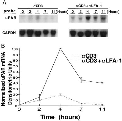
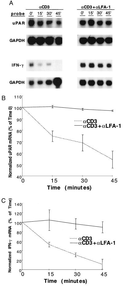
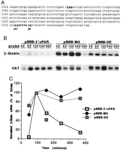
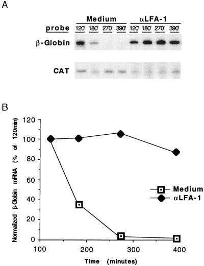
As an adhesion receptor, the beta2 integrin lymphocyte function-associated antigen-1 (LFA-1) contributes a strong adhesive force to promote T lymphocyte recirculation and interaction with antigen-presenting cells. As a signaling molecule, LFA-1-mediates transmembrane signaling, which leads to the generation of second messengers and costimulation resulting in T cell activation. We recently have demonstrated that, in costimulatory fashion, LFA-1 activation promotes the induction of T cell membrane urokinase plasminogen activator receptor (uPAR) and that this induced uPAR is functional. To investigate the mechanism(s) of this induction, we used the RNA polymerase II inhibitor 5, 6-dichloro-1-beta-D-ribobenzimidazole and determined that uPAR mRNA degradation is delayed by LFA-1 activation. Cloning of the wild-type, deleted and mutated 3'-untranslated region of the uPAR cDNA into a serum-inducible rabbit beta-globin cDNA reporter construct revealed that the AU-rich elements and, in particular the nonameric UUAUUUAUU sequence, are crucial cis-acting elements in uPAR mRNA degradation. Experiments in which Jurkat T cells were transfected with reporter constructs demonstrated that LFA-1 engagement was able to stabilize the unstable reporter mRNA containing the uPAR 3'-untranslated region. Our study reveals a consequence of adhesion receptor-mediated signaling in T cells, which is potentially important in the regulation of T cell activation, including production of cytokines and expression of proto-oncogenes, many of which are controlled through 3' AU-rich elements.


Figures

Figure 1
Figure 1
LFA-1-mediated costimulatory effects on uPAR mRNA expression. (A) Jurkat cells were panned with the noted antibody combinations, and total RNA was harvested for Northern analysis at the indicated time points. (B) Curves display relative GAPDH-normalized, uPAR mRNA densitometric units and are representative of four separate experiments, with mean values ± SE.
Figure 2
Figure 2
Effect of LFA-1 engagement on T cell activation mRNA degradation. (A) Jurkat cells (for uPAR) or peripheral T cells (for IFN-γ) were treated with anti-CD3 or anti-CD3 plus anti-LFA-1 antibodies, panned, and incubated × 3 h at 37°C, after which transcription was arrested by the addition of DRB (0.2 mM). Antibody concentrations were as per Materials and Methods section, except that anti-CD3 0.5 μg/107 cells (five times standard protocol) were used to generate higher uPAR mRNA levels with anti-CD3 alone, allowing more easily interpretable decay curves. Total RNA was harvested at the indicated time points and used for uPAR, IFN-γ, and GAPDH Northern blots. Data displayed are representative of results from five separate experiments. Northern signals were densitometrically analyzed and displayed as % of maximal (time 0) GAPDH-normalized, densitometric units with mean values ± SE, for uPAR (B) and IFN-γ (C).
Figure 3
Figure 3
Effect of uPAR 3′ UTR on degradation of a stable mRNA. (A) Jurkat cells were cotransfected with the pEF-BOS-CAT normalization construct and either pBBB or pBBB 3′ uPAR. Cells were fetal bovine serum-stimulated after a 24 h serum starvation, after which poly(A)+ RNA was isolated at the indicated time points for sequential β-globin and CAT Northern analyses. The larger β-globin mRNA size in the pBBB 3′ uPAR samples reflects the additional 263 nt of 3′ uPAR cloned into the pBBB construct. Results are representative of six separate experiments. (B) β-globin Northern signals from A were densitometrically analyzed and normalized to CAT signals. Data represent percentage of maximal (90 min) counts.
Figure 4
Figure 4
Degradation of β-globin mRNA containing wild-type or mutant uPAR 3′ UTR. (A) The 3′ uPAR cDNA, with stop codon (taa) bold and underlined. Sequence 1084–1347 was subcloned into pBBB to generate pBBB-3′ uPAR. AU-rich region is italicized and deleted in pBBB-DE. The nonameric degradation motif is italicized and bolded. The underlined bases (t) in this sequence were mutated to g and c, as indicated, to generate pBBB-MU. (B) Jurkat cells were cotransfected with pEF-BOS-CAT and either pBBB 3′ uPAR, pBBB-MU, or pBBB-DE. Cells were fetal bovine serum-stimulated after a 24-h serum starvation, after which total RNA was harvested at the indicated time points for ribonuclease protection assay. Findings were similar in three separate experiments. (C) β-globin RPA signals from B were densitometrically analyzed and normalized to CAT signals. Isolated data point in the pBBB-DE curve (150 min) represents an aberrant experimental sample, not reproduced in other similar experiments.
Figure 5
Figure 5
LFA-1 engagement stabilizes β-globin mRNA containing the uPAR 3′ ARE. (A) Jurkat T cells were cotransfected with pBBB-3′ uPAR and pEF-BOS-CAT. Cells were serum starved for 24 h, the last 1 h of which included panning with anti-LFA-1 mAbs. Cells were then fetal bovine serum-stimulated at 37°C and total RNA harvested at the indicated time points for RPA. Three separate experiments yielded identical results. (B) β-Globin RPA signals from A were densitometrically analyzed and normalized to CAT signals.

Similar articles

Cited by

References

    1. Hynes R O. Cell. 1992;69:11–25. - PubMed
    1. Schwartz R H. Cell. 1992;71:1065–1068. - PubMed
    1. Fan S-T, Brian A A, Lollo B A, Mackman N, Shen N L, Edington T S. Cell Immunol. 1993;148:48–59. - PubMed
    1. Damle N K, Klussman K, Leytze G, Myrdal S, Aruffo A, Ledbetter J A, Linsley P S. J Immunol. 1994;152:2686– 2697. - PubMed
    1. Van Seventer G A, Shimizu Y, Horgan K J, Shaw S. J Immunol. 1990;144:4579–4586. - PubMed

Publication types

MeSH terms

LinkOut - more resources