. 1998 May 26;95(11):6320-5.
doi: 10.1073/pnas.95.11.6320.

Murine Nkg2d and Cd94 are clustered within the natural killer complex and are expressed independently in natural killer cells

Affiliations

Murine Nkg2d and Cd94 are clustered within the natural killer complex and are expressed independently in natural killer cells

E L Ho et al. Proc Natl Acad Sci U S A. .

Abstract

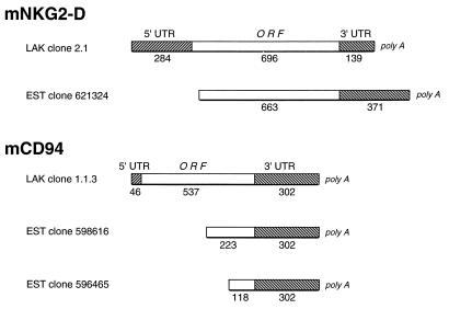
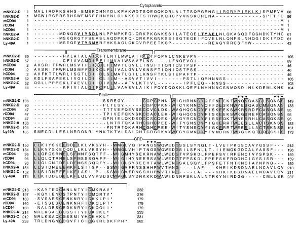
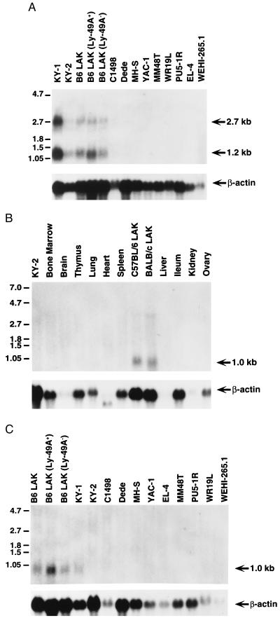
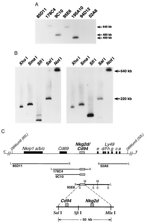
Natural killer (NK) cells express C-type lectin-like receptors, encoded in the NK gene complex, that interact with major histocompatibility complex class I and either inhibit or activate functional activity. Human NK cells express heterodimers consisting of CD94 and NKG2 family molecules, whereas murine NK cells express homodimers belonging to the Ly-49 family. The corresponding orthologues for other species, however, have not been described. In this report, we used probes derived from the expressed sequence tag database to clone C57BL/6-derived cDNAs homologous to human NKG2-D and CD94. Among normal tissues, murine NKG2-D and CD94 transcripts are highly expressed only in activated NK cells, including both Ly-49A+ and Ly-49A- subpopulations. Additionally, mNKG2-D is expressed in murine NK cell clones KY-1 and KY-2, whereas mCD94 expression is observed only in KY-1 cells but not KY-2. Last, we have finely mapped the physical location of the Cd94 (centromeric) and Nkg2d (telomeric) genes between Cd69 and the Ly49 cluster in the NK complex. Thus, these data indicate the expanding complexity of the NK complex and the corresponding repertoire of C-type lectin-like receptors on murine NK cells.


Figures

Figure 1
Figure 1
Schematic diagram depicting the relationship between the murine LAK cDNA clones for mNKG2-D (Upper) and mCD94 (Lower) and the corresponding EST clones. Hatched boxes, the 5′ and 3′ UTRs; open boxes, coding sequences of the ORFs. The numbers below each section indicate length in nucleotides.
Figure 2
Figure 2
Amino acid alignment of the C type lectins in the CD94/NKG2 family and Ly-49A. Brackets indicate boundaries for the cytoplasmic, transmembrane, stalk, and carbohydrate recognition domains (CRD). Regions of identity are shaded for emphasis and include tryptophan and cysteine residues that are conserved in all C-type lectins as well as spacing (48, 49). Consensus ITIM (boldface type underlined), charged residues in transmembrane domain in mNKG2-D and mCD94 (circled), stop codon (∗), and potential N-linked glycosylation sites (♦♦♦) are shown. The 13-amino acid sequence present in the cytoplasmic domain of mNKG2-D but not hNKG2-D is doubly underlined.
Figure 3
Figure 3
Northern blot analyses of mNKG2-D and mCD94 transcripts. (A) Ten micrograms of total cellular RNA from the indicated cells and cell lines was hybridized with full-length mNKG2-D cDNA. (B) Twenty micrograms of total cellular RNA from the indicated organs and cultured cells were hybridized with full-length mCD94 cDNA. (C) Twenty micrograms of total cellular RNA from the indicated LAK cell populations and cell lines were hybridized with the mCD94 cDNA. All of the blots were rehybridized with a human β-actin probe to control for RNA quality and content, as shown below.
Figure 4
Figure 4
Mapping of murine Nkg2d and Cd94 within the NKC. (A) Southern blot analysis of YACs previously shown to form a contig spanning an ≈2.0-megabase region of the murine NKC on chromosome 6 (39), schematically represented in C. This blot was hybridized with both mCD94 (shown) and mNKG2-D (not shown) full-length cDNA probes; identical patterns were observed. (B) Southern blot analysis of YAC 95E6 digested with the indicated enzymes and hybridized with mNKG2-D (Left) or mCD94 (Right) cDNA probes. The murine Nkg2d and Cd94 loci are discriminated on SfiI digestion of YAC 95E6. (C) Schematic representation of the murine Nkg2d and Cd94 loci (hatched boxes) positioned between Cd69 and the Ly49 cluster (solid boxes). The positions of each YAC previously aligned within the NKC contig are shown below (solid lines). Where known, the right (R) and left (L) ends of each YAC are indicated. SalI (S) and Mlu I (M) sites present in YAC 95E6 are shown for reference. An expanded region of YAC 95E6 representing the ≈50-kb SalI–Mlu I fragment containing both loci is shown at the bottom. The exact position of the SfiI site separating the Cd94 and Nkg2d loci and the precise location of the individual genes on the fragment have not been determined.

References

    1. Trinchieri G. Adv Immunol. 1989;47:187–376. - PMC - PubMed
    1. Bancroft G J. Curr Opin Immunol. 1993;5:503–510. - PubMed
    1. Yokoyama W M. Proc Natl Acad Sci USA. 1995;92:3081–3085. - PMC - PubMed
    1. Colonna M. Curr Opin Immunol. 1996;8:101–107. - PubMed
    1. Brown M G, Scalzo A A, Matsumoto K, Yokoyama W M. Immunol Rev. 1997;155:53–65. - PubMed

Publication types

Associated data