. 1998 May 26;95(11):6343-8.
doi: 10.1073/pnas.95.11.6343.

A human fibrosarcoma inhibits systemic angiogenesis and the growth of experimental metastases via thrombospondin-1

Affiliations

A human fibrosarcoma inhibits systemic angiogenesis and the growth of experimental metastases via thrombospondin-1

O V Volpert et al. Proc Natl Acad Sci U S A. .

Abstract

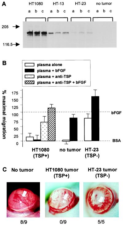
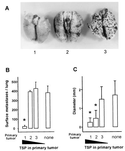
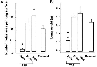
Concomitant tumor resistance refers to the ability of some large primary tumors to hold smaller tumors in check, preventing their progressive growth. Here, we demonstrate this phenomenon with a human tumor growing in a nude mouse and show that it is caused by secretion by the tumor of the inhibitor of angiogenesis, thrombospondin-1. When growing subcutaneously, the human fibrosarcoma line HT1080 induced concomitant tumor resistance, preventing the growth of experimental B16/F10 melanoma metastases in the lung. Resistance was due to the production by the tumor cells themselves of high levels of thrombospondin-1, which was present at inhibitory levels in the plasma of tumor-bearing animals who become unable to mount an angiogenic response in their corneas. Animals carrying tumors formed by antisense-derived subclones of HT1080 that secreted low or no thrombospondin had weak or no ability to control the growth of lung metastases. Although purified human platelet thrombospondin-1 had no effect on the growth of melanoma cells in vitro, when injected into mice it was able to halt the growth of their experimental metastases, providing clear evidence of the efficacy of thrombospondin-1 as an anti-tumor agent.


Figures

Figure 1
Figure 1
Systemic effects of tumors producing TSP-1. (A) Nude mice bearing tumors derived from cells producing various amounts of TSP-1 were examined for human TSP-1 levels present in plasma of tumor-bearing animals by Western analysis. Letters denote individual animals and cells that gave rise to tumors are indicated above each set of samples. (B) In vitro angiogenic activity of the plasma of tumor-bearing animals. Plasma of animals bearing no tumor or tumors derived from HT1080, producing high levels of TSP-1, or from HT-23, producing negligible levels of TSP-1, were tested for the ability to inhibit the migration of capillary endothelial cells toward bFGF in the presence and in the absence of neutralizing anti-TSP-1 antibodies. Data is presented as percent (%) of maximum migration, with 0% set to background levels (BSA) and 100% to the migration toward bFGF. The 100% varied in these experiments; between 43 and 87 cells migrated per ten high powered fields. When tested alone the antibodies did not influence basal or induced migration. (C) Ability of tumor-bearing animals to mount an angiogenic response. Hydron-sucralfate pellets containing bFGF were implanted in the avascular cornea of untreated mice (Left) or of mice bearing tumors formed by parental HT1080 (Middle) or by HT-23 (Right). Note vigorous vessel ingrowth on Left and Right only. Total positive responses/number of implants are tallied below pictures. White arrow indicates position of pellet.
Figure 2
Figure 2
Experimental metastases in tumor-bearing animals. Groups of four animals with no tumor (none) or bearing 5-mm diameter tumors derived from parental HT1080 that produced high levels of TSP- 1 (1), antisense clone HT-13 that produced intermediate TSP-1 levels (2), or antisense clone HT-23 that produced low levels of TSP-1 (3) and received tail vein injections of 106 melanotic B16-F10(P) cells, and 14 days later, lungs were harvested and photographed (A), metastases counted (B), and their average size measured (C). Errors are SEM. ∗Indicates significant difference from nontumor-bearing animals (none) P < 0.01.
Figure 3
Figure 3
Experimental metastases in animals treated with exogenous TSP-1. Animals received an injection of amelanotic B16F10 cells on day 1 and thereafter were divided into groups of four which were treated from day 2 to day 23 twice a day with i.p. injections of 10 mg/kg of purified TSP-1 (daily TSP) or an equal volume of PBS vehicle (PBS). One group was treated with TSP-1 only on the day before and the day after the B16F10 injection (Pulse of TSP). A second group was treated with TSP-1 until day 23 and then remained untreated for 14 days (Reversal). Lungs were harvested and metastases counted (A) and lung weight determined (B). Errors show SEM. ∗Indicates significant difference from PBS control, P < 0.05.

Similar articles

Cited by

References

    1. Prehn R T. Cancer Res. 1991;51:2–4. - PubMed
    1. Prehn R T. Cancer Res. 1993;53:3266–3269. - PubMed
    1. O’Reilly M S, Holmgren L, Shing Y, Chen C, Rosenthal R A, Moses M, Lane W S, Cao Y, Sage E H, Folkman J. Cell. 1994;79:315–328. - PubMed
    1. Holmgren L, O’Reilly M S, Folkman J. Nat Med. 1995;1:149–153. - PubMed
    1. Chen C, Parangi S, Tolentino M J, Folkman J. Cancer Res. 1995;55:4230–4233. - PubMed

Publication types

MeSH terms

Substances

LinkOut - more resources