. 1998 May 26;95(11):6349-54.
doi: 10.1073/pnas.95.11.6349.

In vivo detection and imaging of phosphatidylserine expression during programmed cell death

Affiliations

In vivo detection and imaging of phosphatidylserine expression during programmed cell death

F G Blankenberg et al. Proc Natl Acad Sci U S A. .

Abstract

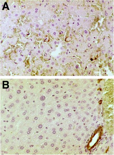
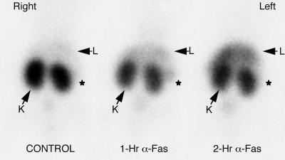
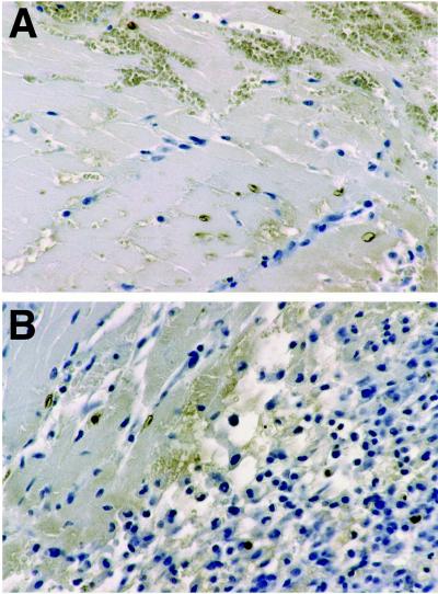
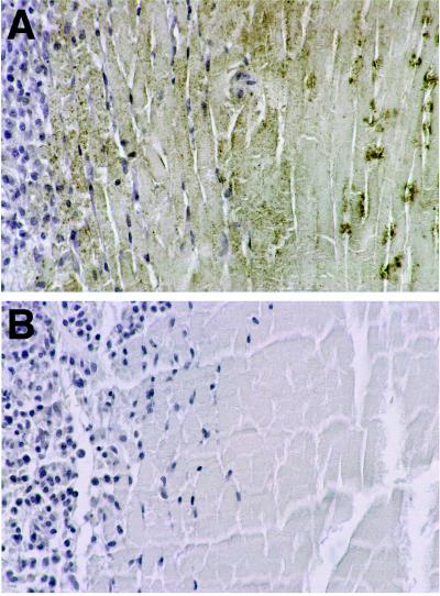
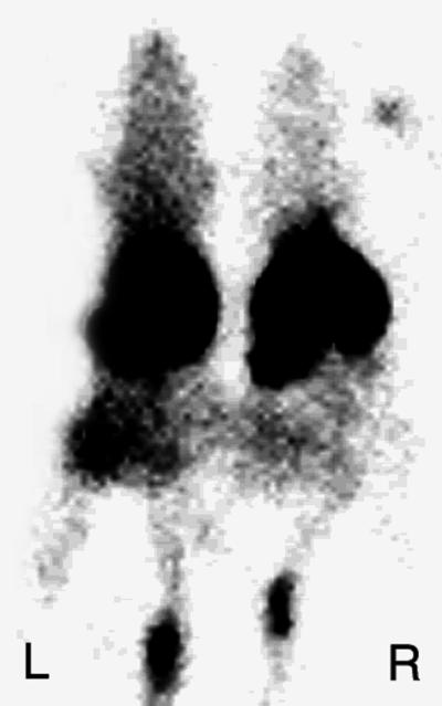
One of the earliest events in programmed cell death is the externalization of phosphatidylserine, a membrane phospholipid normally restricted to the inner leaflet of the lipid bilayer. Annexin V, an endogenous human protein with a high affinity for membrane bound phosphatidylserine, can be used in vitro to detect apoptosis before other well described morphologic or nuclear changes associated with programmed cell death. We tested the ability of exogenously administered radiolabeled annexin V to concentrate at sites of apoptotic cell death in vivo. After derivatization with hydrazinonicotinamide, annexin V was radiolabeled with technetium 99m. In vivo localization of technetium 99m hydrazinonicotinamide-annexin V was tested in three models: fuminant hepatic apoptosis induced by anti-Fas antibody injection in BALB/c mice; acute rejection in ACI rats with transplanted heterotopic PVG cardiac allografts; and cyclophosphamide treatment of transplanted 38C13 murine B cell lymphomas. External radionuclide imaging showed a two- to sixfold increase in the uptake of radiolabeled annexin V at sites of apoptosis in all three models. Immunohistochemical staining of cardiac allografts for exogenously administered annexin V revealed intense staining of numerous myocytes at the periphery of mononuclear infiltrates of which only a few demonstrated positive apoptotic nuclei by the terminal deoxynucleotidyltransferase-mediated UTP end labeling method. These results suggest that radiolabeled annexin V can be used in vivo as a noninvasive means to detect and serially image tissues and organs undergoing programmed cell death.


Figures

Figure 1
Figure 1
Histologic sections of murine liver immunostained for exogenously administered human annexin V. (A) Two hours after anti-Fas antibody treatment there is extensive apoptotic nuclear change, slight cytoplasmic retraction, and interstitial hemorrhage. Annexin V staining (brown immunostaining product) is focally present at the cytoplasmic border of apoptotic hepatocytes. (B) No hepatocyte staining was observed in untreated mice. Staining of bile duct epithelium was caused by antibody cross-reactivity because it also was seen in the absence of exogenously administered human annexin V (data not shown). (Diaminobenzidine immunostain with hematoxylin counterstain, ×40 objective magnification.)
Figure 2
Figure 2
Imaging Fas-mediated fulminant hepatic apoptosis with radiolabeled annexin V. One hour after injection of 150 μCi of radiopharmaceutical (50 μg/kg of protein) mice were imaged in the prone anterior projection. There was a progressive increase in 99mTc annexin V uptake of the liver of mice at 1 and 2 hr after anti-Fas antibody injection. Liver activity (L) was 111% and 239% above control values in the 1- and 2-hr mice, respectively, as shown by region of interest image analysis. Kidney activity (K) was 70% and 64% below control values in the 1- and 2-hr mice, respectively. Splenic activity (*) was 168% and 45% above control values in the 1- and 2-hr mice, respectively.
Figure 3
Figure 3
Imaging cardiac allograft rejection with radiolabeled annexin V. Representative images of abdominal cardiac syngeneic ACI isograft and PVG allograft in ACI host rats 5 days after transplantation. Rats were imaged in the prone anterior projection 1 hr after injection with 900 μCi of 99mTc-annexin V. Location of transplanted hearts are marked by arrows. Intense uptake of 99mTc HYNIC-annexin V was observed in the cardiac allograft animal (Right) as compared with the lack of visualization of the syngeneic cardiac isograft (Left).
Figure 4
Figure 4
Staining for apoptotic nuclei in allografted rodent heart 5 days after transplantation. (A) TUNEL staining showing apoptotic nuclei and fragments in some myocytes bordering areas of necrosis (myocytes without visible nuclei in upper half of field). (B) TUNEL positive nuclei and fragments within inflammatory infiltrate (right half of field) and in some myocytes bordering regions of inflammation. (Diaminobenzidine immunostain with hematoxylin counterstain, ×40 objective magnification.)
Figure 5
Figure 5
Immunostaining for exogenously administered human annexin V in allografted rodent heart 5 days after transplantation. (A) Immunostaining with an antibody to human annexin V shows dense, granular staining of apoptotic myocytes at the periphery of the inflammatory infiltrate. Staining of necrotic myocytes (myocytes without visible nuclei) was clumped heavily and central. (B) Immunostaining of area similar to that shown in A omitting the primary antibody, shows no reaction product. (Diaminobenzidine immunostain with hematoxylin counterstain, ×40 objective magnification.)
Figure 6
Figure 6
Imaging treated murine lymphoma with radiolabeled annexin V. CH3.HeN mice 14 days after implantation of 38C13 murine B cell lymphoma s.c. into the left flank were treated with 100 mg/kg of cyclophosphamide injected i.p. Twenty hours after treatment mice were injected with 150 μCi of 99mTc HYNIC annexin V (50 μg/kg of protein). One hour after administration of 99mTc HYNIC annexin V mice were imaged in the prone anterior projection. Treated tumor demonstrated uptakes of 363% and 454% above control seen by region of interest analysis and biodistribution assay, respectively. Control tumor weight = 1.29 grams, treated tumor weight = 0.82 grams. L, left; R, right.

Similar articles

Cited by

References

    1. Thompson B C. Science. 1995;267:1456–1462. - PubMed
    1. Steller H. Science. 1995;267:1445–1449. - PubMed
    1. Darzynkiewicz Z. J Cell Biochem. 1995;58:151–159. - PubMed
    1. Kerr J F, Wyllie A H, Currie A R. Brit J Cancer. 1972;26:239–257. - PMC - PubMed
    1. Blankenberg F G, Storrs R W, Naumovski L, Goralski T, Spielman D. Blood. 1996;87:1951–1956. - PubMed

Publication types

MeSH terms