. 1998 May 26;95(11):6367-72.
doi: 10.1073/pnas.95.11.6367.

Angiostatin gene transfer: inhibition of tumor growth in vivo by blockage of endothelial cell proliferation associated with a mitosis arrest

Affiliations

Angiostatin gene transfer: inhibition of tumor growth in vivo by blockage of endothelial cell proliferation associated with a mitosis arrest

F Griscelli et al. Proc Natl Acad Sci U S A. .

Abstract

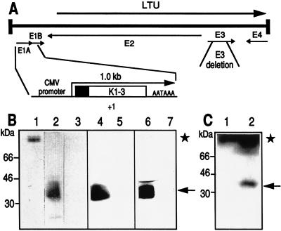
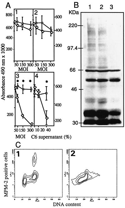
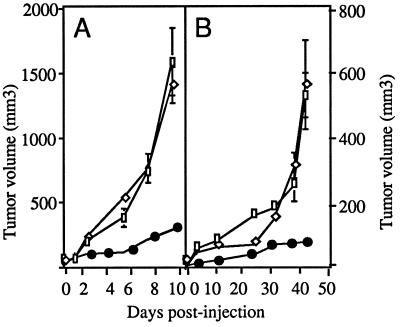
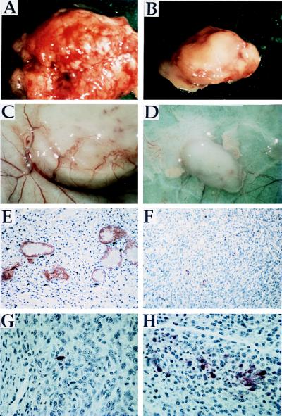
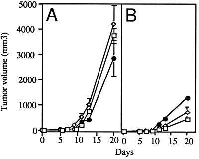
The antitumoral effects that follow the local delivery of the N-terminal fragment of human plasminogen (angiostatin K3) have been studied in two xenograft murine models. Angiostatin delivery was achieved by a defective adenovirus expressing a secretable angiostatin K3 molecule from the cytomegalovirus promoter (AdK3). In in vitro studies, AdK3 selectively inhibited endothelial cell proliferation and disrupted the G2/M transition induced by M-phase-promoting factors. AdK3-infected endothelial cells showed a marked mitosis arrest that correlated with the down-regulation of the M-phase phosphoproteins. A single intratumoral injection of AdK3 into preestablished rat C6 glioma or human MDA-MB-231 breast carcinoma grown in athymic mice was followed by a significant arrest of tumor growth, which was associated with a suppression of neovascularization within and at the vicinity of the tumors. AdK3 therapy also induced a 10-fold increase in apoptotic tumor cells as compared with a control adenovirus. Furthermore, we showed that systemic injection of AdK3 delayed C6 tumor establishment and growth, confirming that angiostatin can function in a paracrin manner. Our data support the concept that targeted antiangiogenesis, using adenovirus-mediated gene transfer, represents a promising alternative strategy for delivering antiangiogenic factors as their bolus injections present unsolved pharmacological problems.


Figures

Figure 1
Figure 1
Recombinant adenoviruses. (A) The Ad5 genome is a 36-kilobase long chromosome. Viruses AdK3 and AdCO1 were derived by a lethal deletion of the E1 genes (nucleotides 382 to 3446); they also carry a nonlethal 1.9-kilobase XbaI deletion within region E3 (for a review, see ref. 23). The angiostatin expression cassette is shown under the Ad5 chromosome. The plasminogen secretion signal is represented by a blackened box; +1 refers to the CMV-driven transcription start; AATAAA refers to the SV40 late polyadenylation signal. (B) Analysis of angiostatin secretion from infected cells. One hundred nanograms of human Plg (lane 1), culture medium from HMEC-1 infected with AdK3 (lane 2) or AdCO1 (lane 3), C6 infected with AdK3 (lane 4) or AdCO1 (lane 5), and MDA-MB-231 infected with AdK3 (lane 6) or AdCO1 (lane 7) were submitted to Western blot analysis. (C) Immunodetection of angiostatin within C6 tumor extracts; Tumors were established in nude mice and received 109 pfu of AdCO1 (lane 1) or AdK3 (lane 2), and Western blot analysis was performed 10 days p.i. The signal corresponding to angiostatin (36–38 kDa) and Plg (92 kDa) are indicated (arrow and asterisk, respectively).
Figure 2
Figure 2
(A) Inhibition of endothelial cell proliferation. C6 (1), MDA-MB-231 (2), and HMEC-1 (3) were injected with AdK3 (◊) or Ad-CO1 (□). HMEC-1 cells (4) cultured with the supernatant from AdK3- (◊) or AdCO1-infected C6 glioma cells (□). (B) Detection of MPM-2 phosphoepitope in HMEC-1 cells. Mock-infected cells (1), AdCO1-infected cells (2), and AdK3-infected cells (3). (C) MPM-2 epitope were detected in HMEC-1 infected with AdCO1 (1) or AdK3 (2) by indirect immunostaining and DNA content by propidium iodide staining, and quantified by flow cytometry (see Materials and Methods). A Student’s t test was used for statistical analysis.
Figure 3
Figure 3
AdK3 inhibits tumor growth. C6 glioma (A) and MDA-MB-231 carcinoma (B) were s.c. implanted into athymic mice (see Materials and Methods). When the tumor had reached a volume of 20 mm3 (day 0), mice received an intratumoral injection of PBS (□), 109 pfu of AdK3 (•), or AdCO1 (◊). Mean values are represented with their standard deviations.
Figure 4
Figure 4
AdK3 inhibits C6 tumor growth and angiogenesis. Tumors from AdCO1-treated (A) and AdK3-treated groups (B) are shown 10 days p.i. The extent of vascularization at the periphery of a representative tumor injected with AdCO1 (C) or AdK3 (D) is shown at day 5 p.i. Paraffin-embeded C6 sections from an AdCO1-injected (E) or AdK3-injected tumor (F) were submitted to vWF-immunostaining at day 10 p.i. The proportion of apoptotic cells was detected by the TUNEL method within sections from an AdCO1-injected (G) or AdK3-injected tumor (H). The same magnification was used for AdCO1- and AdK3-injected tumors.
Figure 5
Figure 5
Dose-dependent effect of AdK3. C6 cells were infected in vitro, 24 hr with AdCO1 (A) or AdK3 (B), and mixed at a ratio of 1 (□), 1:2 (◊), and 1:4 (•) with noninfected C6 cells before implantation into athymic mice (n = 6). Tumor volumes were measured during 20 days. Mean values are represented with their standard deviations.

Similar articles

Cited by

References

    1. Folkman J. J Natl Cancer Inst. 1990;82:4–6. - PubMed
    1. Folkman J. Nat Med. 1995;1:27–31. - PubMed
    1. Hanahan D, Folkman J. Cell. 1996;86:353–364. - PubMed
    1. Hori A. Cancer Res. 1991;51:6180–6184. - PubMed
    1. Kim K J. Nature (London) 1993;362:841–844. - PubMed

Publication types

LinkOut - more resources