. 1998 May 26;95(11):6373-8.
doi: 10.1073/pnas.95.11.6373.

Abnormal transport along the lysosomal pathway in mucolipidosis, type IV disease

Affiliations

Abnormal transport along the lysosomal pathway in mucolipidosis, type IV disease

C S Chen et al. Proc Natl Acad Sci U S A. .

Abstract

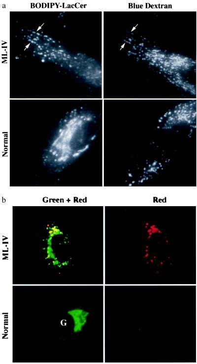
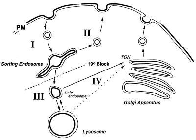
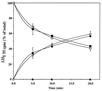
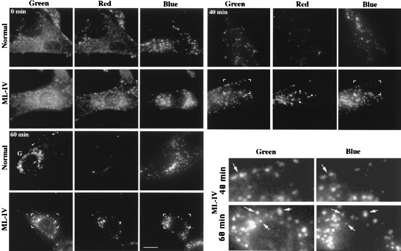
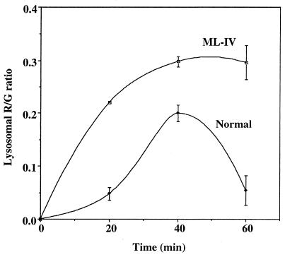
Mucolipidosis, type IV (ML-IV) is an autosomal recessive storage disease that is characterized by lysosomal accumulation of sphingolipids, phospholipids, and acid mucopolysaccharides. Unlike most other storage diseases, the lysosomal hydrolases participating in the catabolism of the stored molecules appear to be normal. In the present study, we examined the hypothesis that the ML-IV phenotype might arise from abnormal transport along the lysosomal pathway. By using various markers for endocytosis, we found that plasma membrane internalization and recycling were nearly identical in ML-IV and normal fibroblasts. A fluorescent analog of lactosylceramide (LacCer) was used to study plasma membrane lipid internalization and subsequent transport. Lipid internalization at 19 degreesC was similar in both cell types; however, 40-60 min after raising the temperature to 37 degreesC, the fluorescent lipid accumulated in the lysosomes of ML-IV cells but was mainly concentrated at the Golgi complex of normal fibroblasts. Biochemical studies demonstrated that at these time points, hydrolysis of the lipid analog was minimal ( approximately 7%) in both cell types. A fluorescence ratio imaging assay was developed to monitor accumulation of fluorescent LacCer in the lysosomes and showed that the apparent concentration of the lipid increased more rapidly and to a greater extent in ML-IV cells than in normal fibroblasts. By 60 min, LacCer apparently decreased in the lysosomes of normal fibroblasts but not in ML-IV cells, suggesting that lipid efflux from the lysosomes was also impaired. These results demonstrate that there is a defect in ML-IV fibroblasts that affects membrane sorting and/or late steps of endocytosis.


Figures

Figure 1
Figure 1
Lysosomal accumulation of C5-DMB-LacCer in ML-IV cells. Normal and ML-IV fibroblasts were incubated overnight with a blue fluorescent dextran to label the lysosomes and subsequently pulse-labeled with C5-DMB-LacCer (see text). (a) In ML-IV cells, many of the punctate structures labeled with the fluorescent lipid colocalized with the dextran-stained lysosomes (e.g., at arrows), but in normal fibroblasts most of the fluorescent lipid was observed at the Golgi complex (G), with little present in the lysosomes. (b) Observations of BODIPY fluorescence in different regions of the spectrum also demonstrated a shift in BODIPY fluorescence toward red wavelengths in ML-IV (but not in normal) fibroblasts.
Figure 2
Figure 2
Vesicular transport along the lysosomal pathway. Pathway I, transport from the plasma membrane (PM) to “sorting endosomes.” The latter is experimentally defined by the use of a 19°C temperature block. Pathway II, “recycling” from sorting endosomes back to the plasma membrane. Pathway III, transport from sorting endosomes to lysosomes, was studied by measuring the concentration-dependent spectral changes in fluorescence as a BODIPY-lipid accumulated in the lysosomes. Pathway IV, alternative pathways for lipid transport to the Golgi complex. TGN, trans-Golgi network.
Figure 3
Figure 3
Recycling kinetics in normal vs. ML-IV cells. Normal (•, ▪, and ▴) and ML-IV (○, □, and ▵) fibroblasts were incubated with 125I-Tfn for 60 min at 19°C, washed, and “acid-stripped” to remove any labeled Tfn at the plasma membrane. The cells were then warmed to 37°C for various times after which the amount of cell-associated 125I-Tfn (surface, ▴ and ▵; intracellular, ▪ and □) and 125I-Tfn released into the medium (• and ○) was determined.
Figure 4
Figure 4
Transport of C5-DMB-LacCer from sorting endosomes to lysosomes in normal vs. ML-IV cells. Normal or ML-IV cells were preincubated with blue dextran to label the lysosomes. The cells were then incubated for 1 hr at 19°C with 5 μM C5-DMB-LacCer/defatted BSA, washed, back-exchanged with 5% defatted BSA, and further incubated for the indicated times at 37°C. Samples were photographed in the blue (dextran), green (BODIPY), and red (BODIPY) regions of the spectrum. (Inset) Bracketed regions show that some of the BODIPY-labeled vesicles are coincident with the lysosomes of ML-IV cells (e.g., at arrows). Also, note the labeling of the Golgi complex (G) in normal but not ML-IV fibroblasts at 60 min. (Bar = 10 μm.)
Figure 5
Figure 5
Temporal changes in accumulation of C5-DMB-LacCer at the lysosomes. Normal and ML-IV cells were incubated with blue dextran and C5-DMB-LacCer/defatted BSA as in Fig. 4. Images were acquired at each time point and the R/G ratio of individual lysosomes was quantified. Attempts to quantify the R/G ratio at earlier time points were not reliable because there was often substantial overlap of lysosomes and endosomes in the same region of the cell at these times, making it difficult to generate an appropriate “lysosome mask” for these calculations.

References

    1. Bach G, Zeigler M, Kohn G, Cohen M M. Am J Hum Genet. 1977;29:610–618. - PMC - PubMed
    1. Amir N, Zlotogora J, Bach G. Pediatrics. 1987;79:953–959. - PubMed
    1. Bach G, Zeigler M, Bargal R. In: Advances in Jewish Genetic Diseases. Desnick R J, editor. New York: Oxford Univ. Press; 1998. , in press.
    1. Bargal R, Bach G. J Inherited Metab Dis. 1997;20:625–631. - PubMed
    1. Koval M, Pagano R E. J Cell Biol. 1990;111:429–442. - PMC - PubMed

Publication types