

Regulation of the pAD1 sex pheromone response of Enterococcus faecalis by direct interaction between the cAD1 peptide mating signal and the negatively regulating, DNA-binding TraA protein
- PMID: 9600983
- PMCID: PMC27773
- DOI: 10.1073/pnas.95.11.6430
Regulation of the pAD1 sex pheromone response of Enterococcus faecalis by direct interaction between the cAD1 peptide mating signal and the negatively regulating, DNA-binding TraA protein
Abstract
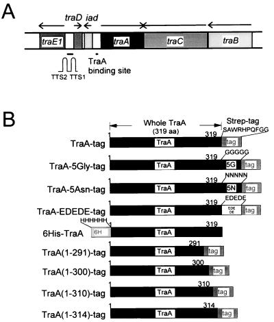
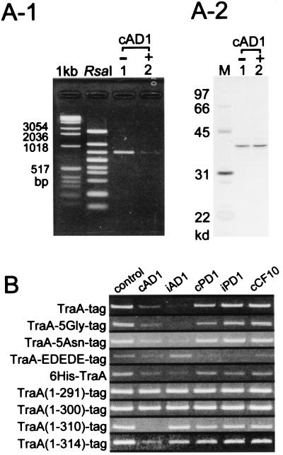
The Enterococcus faecalis conjugative plasmid pAD1 (60 kb) encodes a mating response to the recipient-produced peptide sex pheromone cAD1. The response involves two key plasmid-encoded regulatory proteins: TraE1, which positively regulates all or most structural genes relating to conjugation, and TraA, which binds DNA and negatively regulates expression of traE1. In vitro studies that included development of a DNA-associated protein-tag affinity chromatography technique showed that TraA (37.9 kDa) binds directly to cAD1 near its carboxyl-terminal end and, as a consequence, loses its affinity for DNA. Analyses of genetically modified TraA proteins indicated that truncations within the carboxyl-terminal 9 residues significantly affected the specificity of peptide-directed association/dissociation of DNA. The data support earlier observations that transposon insertions near the 3' end of traA eliminated the ability of cells to respond to cAD1.
Figures
Similar articles
-
Regulation of the pAD1-encoded sex pheromone response in Enterococcus faecalis: expression of the positive regulator TraE1.J Bacteriol. 1993 Feb;175(4):1008-18. doi: 10.1128/jb.175.4.1008-1018.1993. J Bacteriol. 1993. PMID: 8432694 Free PMC article.
-
Regulation of the pAD1-encoded sex pheromone response in Enterococcus faecalis: nucleotide sequence analysis of traA.J Bacteriol. 1992 Mar;174(6):1821-7. doi: 10.1128/jb.174.6.1821-1827.1992. J Bacteriol. 1992. PMID: 1312529 Free PMC article.
-
Regulation of the Enterococcus faecalis pAD1-related sex pheromone response: analyses of traD expression and its role in controlling conjugation functions.Mol Microbiol. 1998 Oct;30(2):381-92. doi: 10.1046/j.1365-2958.1998.01074.x. Mol Microbiol. 1998. PMID: 9791182
-
The peptide pheromone-inducible conjugation system of Enterococcus faecalis plasmid pCF10: cell-cell signalling, gene transfer, complexity and evolution.Philos Trans R Soc Lond B Biol Sci. 2007 Jul 29;362(1483):1185-93. doi: 10.1098/rstb.2007.2043. Philos Trans R Soc Lond B Biol Sci. 2007. PMID: 17360276 Free PMC article. Review.
-
Properties of Enterococcus faecalis plasmid pAD1, a member of a widely disseminated family of pheromone-responding, conjugative, virulence elements encoding cytolysin.Plasmid. 2007 Nov;58(3):205-27. doi: 10.1016/j.plasmid.2007.05.001. Epub 2007 Jun 27. Plasmid. 2007. PMID: 17590438 Review.
Cited by
-
The genes coding for enterocin EJ97 production by Enterococcus faecalis EJ97 are located on a conjugative plasmid.Appl Environ Microbiol. 2003 Mar;69(3):1633-41. doi: 10.1128/AEM.69.3.1633-1641.2003. Appl Environ Microbiol. 2003. PMID: 12620853 Free PMC article.
-
Analysis of the amino acid sequence specificity determinants of the enterococcal cCF10 sex pheromone in interactions with the pheromone-sensing machinery.J Bacteriol. 2007 Feb;189(4):1399-406. doi: 10.1128/JB.01226-06. Epub 2006 Nov 10. J Bacteriol. 2007. PMID: 17098891 Free PMC article.
-
Identification of the cAD1 sex pheromone precursor in Enterococcus faecalis.J Bacteriol. 2002 Apr;184(7):1880-7. doi: 10.1128/JB.184.7.1880-1887.2002. J Bacteriol. 2002. PMID: 11889094 Free PMC article.
-
Identification and characterization of a determinant (eep) on the Enterococcus faecalis chromosome that is involved in production of the peptide sex pheromone cAD1.J Bacteriol. 1999 Oct;181(19):5915-21. doi: 10.1128/JB.181.19.5915-5921.1999. J Bacteriol. 1999. PMID: 10498702 Free PMC article.
-
Functional analysis of TraA, the sex pheromone receptor encoded by pPD1, in a promoter region essential for the mating response in Enterococcus faecalis.J Bacteriol. 2002 Nov;184(22):6343-50. doi: 10.1128/JB.184.22.6343-6350.2002. J Bacteriol. 2002. PMID: 12399504 Free PMC article.
References
-
- Clewell D B. Eur J Clin Microbiol Infect Dis. 1990;9:90–102. - PubMed
Publication types
MeSH terms
Substances
Grants and funding
LinkOut - more resources
Full Text Sources
Molecular Biology Databases
