. 1998 May 26;95(11):6448-53.
doi: 10.1073/pnas.95.11.6448.

Diffusible, nonfibrillar ligands derived from Abeta1-42 are potent central nervous system neurotoxins

Affiliations

Diffusible, nonfibrillar ligands derived from Abeta1-42 are potent central nervous system neurotoxins

M P Lambert et al. Proc Natl Acad Sci U S A. .

Abstract

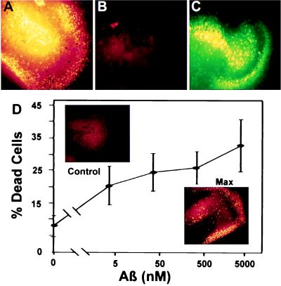
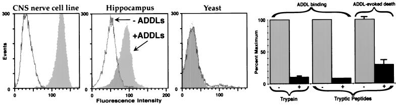
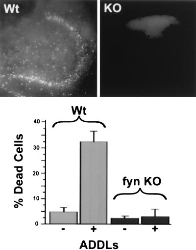
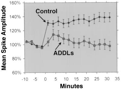
Abeta1-42 is a self-associating peptide whose neurotoxic derivatives are thought to play a role in Alzheimer's pathogenesis. Neurotoxicity of amyloid beta protein (Abeta) has been attributed to its fibrillar forms, but experiments presented here characterize neurotoxins that assemble when fibril formation is inhibited. These neurotoxins comprise small diffusible Abeta oligomers (referred to as ADDLs, for Abeta-derived diffusible ligands), which were found to kill mature neurons in organotypic central nervous system cultures at nanomolar concentrations. At cell surfaces, ADDLs bound to trypsin-sensitive sites and surface-derived tryptic peptides blocked binding and afforded neuroprotection. Germ-line knockout of Fyn, a protein tyrosine kinase linked to apoptosis and elevated in Alzheimer's disease, also was neuroprotective. Remarkably, neurological dysfunction evoked by ADDLs occurred well in advance of cellular degeneration. Without lag, and despite retention of evoked action potentials, ADDLs inhibited hippocampal long-term potentiation, indicating an immediate impact on signal transduction. We hypothesize that impaired synaptic plasticity and associated memory dysfunction during early stage Alzheimer's disease and severe cellular degeneration and dementia during end stage could be caused by the biphasic impact of Abeta-derived diffusible ligands acting upon particular neural signal transduction pathways.


Figures

Figure 1
Figure 1
AFM and gel electrophoresis show that toxic ADDL preparations comprise small, fibril-free oligomers of Aβ1–42. AFM examination of toxic ADDLs shows small globular structures, ≈5–6 nm in size, and a distinct lack of fibrils, consistent with migration of ADDLs as oligomers during gel electrophoresis. (Upper Left) Examination of conventional Aβ preparations by AFM shows primarily large, nondiffusible fibrillar species. (Upper Right) ADDLs imaged by AFM show size and structure consistent with their diffusible nature. (Inset) Native gel of ADDLs made in the cold. Note two major species found at ≈27 and 17 kDa and the absence of large molecular weight species. (Lower Left) Lane W, SDS/PAGE (Western blot) using 6E10 antibody of ADDLs made in the cold. (Lower Right) SDS/PAGE, silver-stained. Lanes: 1, ADDLs made with clusterin; 2, ADDLs made in cold; 3, Centricon 10 retentate of cold-induced ADDLs; 4, Centricon 10 eluate of cold-induced ADDLs. The positions of clusterin monomer, Aβ1–42, and ADDLs are shown.
Figure 2
Figure 2
ADDLs are diffusible, extremely potent CNS neurotoxins. ADDLs in culture medium diffuse through culture support filters and cause extensive cell death in stratum granulosum (DG) and CA3 areas of organotypic hippocampal slice cultures. ADDL toxicity is extensive even at nanomolar doses, but nondiffusing fibrils at 20 μM are not toxic. (A) DG and CA3 area of a hippocampal slice treated for 24 hr with 5 μM ADDLs. Dead cells are highlighted in false yellow color (see Materials and Methods). Up to 40% of the cells in this region die following chronic exposure to ADDLs. (B) DG and CA3 area of another hippocampal slice treated with 20 μM fibrillar Aβ1–42 for 24 hr. No cell death is seen. (C) Live cells in the same area of the slice treated with fibrillar Aβ1–42. (D) ADDLs were added to duplicate mouse hippocampal slices for 24 hr at the indicated concentrations. The Live/Dead assay and image analysis (see Materials and Methods) were used to determine percent dead cells in fields containing the DG and CA3 areas. Data show that even after a 1,000-fold dilution to 5 nM Aβ1–42, ADDLs still kill >20% of the cells, a value greater than half the maximum cell death. (Inset) Comparison of cell death in DG and CA3 observed with vehicle alone (Upper) or with ADDLs at 5 μM (Lower). ADDL concentration was determined by measuring the supernatant protein and subtracting the amount of soluble clusterin present. This formula assumes the amount of clusterin that pellets is negligible.
Figure 3
Figure 3
Cell surface binding of ADDLs is selective and required for toxicity. FACScan shows that ADDL binding is robust in the B103 CNS nerve cell line, lowered in primary hippocampal cells, and completely absent in yeast cells. Consistent with selectivity, trypsinization of cell surfaces blocks subsequent ADDL binding; moreover, cell surface tryptic peptides are antagonists of ADDL binding and toxicity. FACScan assay. (Left) Suspensions of B103 rat neuroblastoma cells (Far Left), primary rat hippocampal cells (Center), and yeast cells (Right, Saccharomyces cerevisieae, log-phase) were incubated with ADDLs for 60 min and then assessed for the presence of bound ADDLs (Materials and Methods). White shows the background fluorescence in absence of ADDLs, gray the increased fluorescence because of addition of ADDLs, and stripes the background level fluorescence in ADDL-treated samples. Bar Graphs (Right) Quantitative comparison shows that ADDL binding in B103 cells is blocked ≈90% by brief trypsinization; furthermore, binding to cells is blocked ≈90% and cell death in the slice assay is blocked ≈75% by the addition of tryptic peptides (see Materials and Methods). Error bars are SEM for four or five replicate samples.
Figure 4
Figure 4
Hippocampal neurotoxicity of ADDLs occurs via a Fyn-dependent pathway. ADDL toxicity is completely blocked in slices from a Fyn knockout (KO) mouse. (Upper) Images of dead cells in the DG and CA3 area of (Left) Fyn wild-type or (Right) Fyn knockout mouse slice exposed to ADDLs for 24 hr. Cell death only occurs with Fyn wild-type genotype. (Lower) Quantitative comparison of cell death in Fyn wild-type and Fyn knockout slices. Error bars are means ± SEM for four to seven slices.
Figure 5
Figure 5
ADDLs block LTP. Incubation of rat hippocampal slices with ADDLs prevents LTP well before any overt signs of cell degeneration. Medial perforant path-granule cell LTP was readily induced in slices from young adult rats (•, see Materials and Methods). In contrast, hippocampal slices exposed to ADDLs for 45 min showed no lasting potentiation (▪), despite a continuing capacity for evoked action potentials.

Similar articles

Cited by

References

    1. Price D L. Annu Rev Neurosci. 1986;9:489–512. - PubMed
    1. Sisodia S S, Price D J. FASEB J. 1995;9:366–370. - PubMed
    1. Yankner B A, Duffy L K, Kirschner D A. Science. 1990;250:279–282. - PubMed
    1. Pike C J, Walencewicz A J, Glabe C G, Cotman C W. Brain Res. 1991;563:311–314. - PubMed
    1. Pike C J, Burdick D, Walencewicz A J, Glabe C G, Cotman C W. J Neurosci. 1993;13:1676–1687. - PMC - PubMed

Publication types

LinkOut - more resources