. 1998 May 26;95(11):6454-9.
doi: 10.1073/pnas.95.11.6454.

Cerebral cortical hyperactivation in response to mental stress in patients with coronary artery disease

Affiliations

Cerebral cortical hyperactivation in response to mental stress in patients with coronary artery disease

R Soufer et al. Proc Natl Acad Sci U S A. .

Abstract

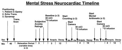
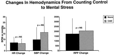
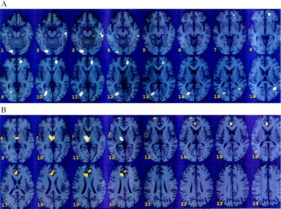
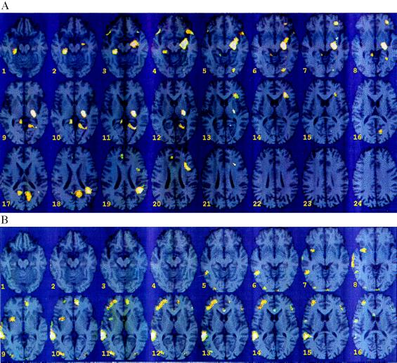
The central nervous system (CNS) effects of mental stress in patients with coronary artery disease (CAD) are unexplored. The present study used positron emission tomography (PET) to measure brain correlates of mental stress induced by an arithmetic serial subtraction task in CAD and healthy subjects. Mental stress resulted in hyperactivation in CAD patients compared with healthy subjects in several brain areas including the left parietal cortex [angular gyrus/parallel sulcus (area 39)], left anterior cingulate (area 32), right visual association cortex (area 18), left fusiform gyrus, and cerebellum. These same regions were activated within the CAD patient group during mental stress versus control conditions. In the group of healthy subjects, activation was significant only in the left inferior frontal gyrus during mental stress compared with counting control. Decreases in blood flow also were produced by mental stress in CAD versus healthy subjects in right thalamus (lateral dorsal, lateral posterior), right superior frontal gyrus (areas 32, 24, and 10), and right middle temporal gyrus (area 21) (in the region of the auditory association cortex). Of particular interest, a subgroup of CAD patients that developed painless myocardial ischemia during mental stress had hyperactivation in the left hippocampus and inferior parietal lobule (area 40), left middle (area 10) and superior frontal gyrus (area 8), temporal pole, and visual association cortex (area 18), and a concomitant decrease in activation observed in the anterior cingulate bilaterally, right middle and superior frontal gyri, and right visual association cortex (area 18) compared with CAD patients without myocardial ischemia. These findings demonstrate an exaggerated cerebral cortical response and exaggerated asymmetry to mental stress in individuals with CAD.


Figures

Figure 1
Figure 1
Timeline of PET mental stress neurocardiac study. Subjects underwent a total of six scans: two baseline, two counting control, and two mental arithmetic. O-15 (30 mCi) was administered during each scan.
Figure 2
Figure 2
Effect of mental stress of heart rate and blood pressure in patients with coronary artery disease (CAD) and normal subjects. There is no significant difference in the increase in heart rate (HR), systolic blood pressure (SBP), and rate pressure product (RPP) from control to mental stress conditions among CAD and normal subjects.
Figure 3
Figure 3
(A) Statistical parametric map overlaid on an MRI template of areas of increased blood flow with mental stress in CAD patients (n = 10) versus healthy subjects (n = 6). There were significant increases in visual association cortex (18) [(Talairach coordinates) x = −26, y = −80, z = 12, z score = 3.32, P < 0.001]; anterior cingulate (32) (x = 14, y = 46, z = 4, z score = 3.08, P < 0.001); cerebellum (x = −22, y = −90, z = −20, z score = 3.00, P < 0.001); left parietal cortex (x = 36, y = −58, z = 36, z score = 2.94, P < 0.002); left fusiform gyrus (x = 52, y = −32, z = −20, z score = 2.88, P < 0.002). (B) Statistical parametric map overlaid on an MRI template of areas of decreased blood flow with mental stress versus counting control in CAD patients (n = 10) versus healthy controls (n = 6). There were significant decreases in thalamus (x = −10, y = −18, z = 16, z score = 4.07, P < 0.001); superior frontal gyrus/cingulate gyrus (32, 24) (x = −6, y = 32, z = 32, z score = 3.29, P < 0.001); superior frontal gyrus (10) (x = −20, y = 58, z = 20, z score −3.15, P < 0.001); right middle temporal gyrus (21) (x = − 60, y = −18, z = 4, z score = 3.08, P < 0.001).
Figure 4
Figure 4
(A) Statistical parametric map overlaid on an MRI template of areas of increased blood flow with mental stress versus counting control in mental stress ischemic CAD patients (n = 3) versus mental stress nonischemic CAD patients (n = 7). There were significant increases in left hippocampus (x = 22, y = −22, z = −8, z score = 4.43, P < 0.001); left parietal cortex (inferior parietal lobule) (40) (x = 40, y = −54, z = 44, z score = 3.80, P < 0.001); left superior frontal gyrus (8) (x = 26, y = 16, z = 52, z score = 3.83, P < 0.001); left middle frontal gyrus (10) (x = 28, y = 36, z = −4, z score = 3.58, P < 0.001); posterior cingulate, corpus callosum (x = 24, y = −62, z = 12, z score = 3.65, P < 0.001); right hippocampus (x = −26, y = −36, z = −24, z score = 3.44, P < 0.001); temporal pole (38) (x = −44, y = 14, z = −16, z score = 3.45, P < 0.001); precuneus (31) (x = −14, y = −58, z = 36, z score = 3.19, P < 0.001); visual association cortex (18) (x = 22, y = −86, z = −12, z score = 3.15, P < 0.001). (B) Statistical parametric map overlaid on an MRI template of areas of decreased blood flow with mental stress versus counting control in mental stress ischemic CAD patients (n = 3) versus mental stress nonischemic CAD patients (n = 7). There were significant decreases in right superior temporal gyrus (22) (x = −58, y = −42, z = 12, z score = 4.84, P < 0.001); right-middle temporal gyrus (37) (x = −46, y = −56, z = −8, z score = 3.60, P < 0.001); left anterior cingulate (x = 8, y = 42, z = 8, z score = 3.91, P < 0.001); right anterior cingulate (24) (x = −8, y = 6, z = 36, z score = 2.89, P = 0.002); visual association cortex (18) (x = −36, y = −88, z = 12, z score = 3.73, P < 0.001); area 19 (x = 34, y = −76, z = 36, z score = 2.66, P < 0.001); right-middle frontal gyrus (10) (x = −24, y = 48, z = 16, z score = 3.61, P < 0.001); insula (x = −32, y = 0, z = 0, z score = 3.45, P < 0.001); precentral (x = −52, y = −6, z = 32, z score = 2.81, P = 0.002); lingual gyrus (x = −16, y = −70, z = 0, z score = 2.39, P = 0.008).

References

    1. Anderson K M, Wilson P W F, Odell P M, Kannel W B. Circulation. 1991;83:356–362. - PubMed
    1. Pepine C D J. Ann Int Med. 1996;124:1006–1007. - PubMed
    1. Rosengren A, Tibblin G, Wilhelmsen L. Am J Cradiol. 1991;68:1171–1175. - PubMed
    1. Ruberman W, Weinblatt E, Goldberg J D, Chaudrey B S. N Engl J Med. 1984;311:552–559. - PubMed
    1. Burg M M, Jain D, Soufer R, Kerns R D, Zaret B L. J Am Coll Cardiol. 1993;22:440–448. - PubMed

Publication types