. 1998 May 26;95(11):6521-5.
doi: 10.1073/pnas.95.11.6521.

The gamma subunit of the Na,K-ATPase induces cation channel activity

Affiliations

The gamma subunit of the Na,K-ATPase induces cation channel activity

N T Minor et al. Proc Natl Acad Sci U S A. .

Abstract

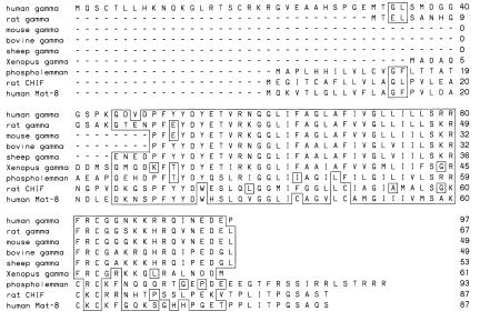
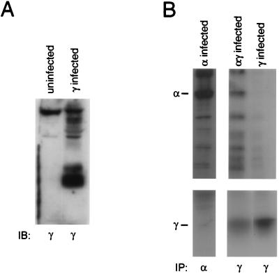
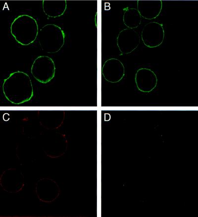
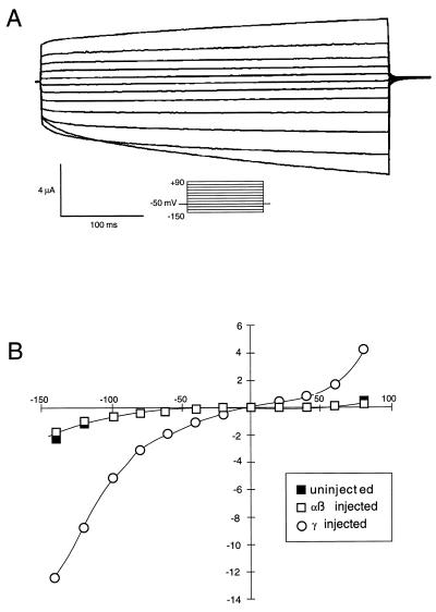
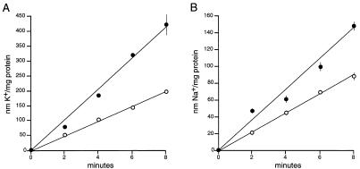
The gamma subunit of the Na,K-ATPase is a hydrophobic protein of approximately 10 kDa. The gamma subunit was expressed in Sf-9 insect cells and Xenopus oocytes to ascertain its role in Na,K-ATPase function. Immunoblotting has shown that the gamma subunit is expressed in Sf-9 cells infected with recombinant baculovirus containing the cDNA for the human gamma subunit. Confocal microscopy demonstrates that the gamma subunit can be delivered to the plasma membrane of Sf-9 cells independently of the other Na,K-ATPase subunits and that gamma colocalizes with alpha1 when these proteins are coexpressed. When Sf-9 cells were coinfected with alpha1 and gamma, antibodies to the gamma subunit were able to coimmunoprecipitate the alpha1 subunit, suggesting that gamma is able to associate with alpha1. The gamma subunit is a member of a family of single-pass transmembrane proteins that induces ion fluxes in Xenopus oocytes. Evidence that the gamma subunit is a functional component was supported by experiments showing gamma-induced cation channel activity when expressed in oocytes and increases in Na+ and K+ uptake when expressed in Sf-9 cells.


Figures

Figure 1
Figure 1
Alignment of the deduced amino acid sequences of the γ subunit of the Na,K-ATPase from various sources to phospholemman, rat channel-inducing factor, and human Mat-8.
Figure 2
Figure 2
Expression of the γ subunit in Sf-9 cells and association with the α1 subunit. (A) An immunoblot of protein from cells infected with baculoviruses containing the cDNA for the human γ subunit or uninfected cells. The blots were probed with a γ specific polyclonal (γ969). (B) An immunoprecipitation of protein from metabolically labeled Sf-9 cells infected with α1, α1γ, or γ baculoviruses. Protein from α1-infected cells was immunoprecipitated with an α1-specific monoclonal (C464–6B). Protein from γ- and α1γ-infected cells was immunoprecipitated with γ969.
Figure 3
Figure 3
Localization of the γ subunit in infected Sf-9 cells. Confocal microscopy was used to determine the location of proteins in cells infected with γ or α1β1γ baculoviruses. (A) Cells infected with γ baculovirus and probed with G17 polyclonal. (BD) Cells coinfected with α1β1γ baculoviruses and probed with both the G17 polyclonal and 5α monoclonal to detect the γ and α subunits. (B) Staining for the γ subunit. (C) Staining for the α1 subunit. (D) an overlay of B and C, showing colocalization of γ and α1 in α1β1γ-infected cells.
Figure 4
Figure 4
Ion currents in Xenopus oocytes injected with γ cRNA. (A) Currents recorded under voltage clamp, from a Xenopus oocyte expressing γ subunits. The membrane was stepped from a holding potential of −50 mV to voltages between −150 and +90 mV, for 400 msec, every 5 sec. The record shown is a typical first set of currents after turning on the voltage clamp. (B) Typical “steady-state” current-voltage relationships from uninjected oocytes, and oocytes expressing α and β subunits, or γ subunits alone, by using a protocol like that shown in A.
Figure 5
Figure 5
Na+ and K+ uptake into γ-infected Sf-9 cells. Cells infected with γ (•) or IAP (○) baculoviruses were depleted of intracellular K+ (A) or Na+ (B) and uptake was measured by using 86Rb or 22Na as tracer, respectively. Bumetanide (100 μM) was included to inhibit bumetanide-sensitive transport. The rate of Na+ or K+ uptake in uninfected cells is approximately 80% of the rate in infected cells (not shown). Each value is the mean with error bars representing the standard error of eight determinations. Similar results were obtained from two different experiments.

References

    1. Noguchi S, Mishina M, Kawamura M, Numa S. FEBS Lett. 1987;225:27–32. - PubMed
    1. Horowitz B, Eakle K A, Scheiner-Bobis G, Randolph G R, Chen C Y, Hitzeman R A, Farley R A. J Biol Chem. 1991;265:4189–4192. - PubMed
    1. Blanco G, Xie Z J, Mercer R W. Proc Natl Acad Sci USA. 1993;90:1824–1828. - PMC - PubMed
    1. Lingrel J B, Orlowski J, Shull M M, Price E M. Prog Nucleic Acid Res Biol. 1990;38:37–89. - PubMed
    1. Forbush B, III, Kaplan J H, Hoffman J F. Biochemistry. 1978;17:3667–3676. - PubMed

Publication types

Substances

LinkOut - more resources