. 1998 May 26;95(11):6560-5.
doi: 10.1073/pnas.95.11.6560.

Precisely full length, circularizable, complementary RNA: an infectious form of potato spindle tuber viroid

Affiliations

Precisely full length, circularizable, complementary RNA: an infectious form of potato spindle tuber viroid

P A Feldstein et al. Proc Natl Acad Sci U S A. .

Abstract

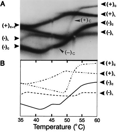
The replication of many viral and subviral pathogens as well as the amplification of certain cellular genes proceeds via a rolling circle mechanism. For potato spindle tuber (PSTVd) and related viroids, the possible role of a circular (-)strand RNA as a template for synthesis of (+)strand progeny is unclear. Infected plants appear to contain only multimeric linear (-)strand RNAs, and attempts to initiate infection with multimeric (-)PSTVd RNAs generally have failed. To examine critically the infectivity of monomeric (-)strand viroid RNAs, we have developed a ribozyme-based expression system for the production of precisely full length (-)strand RNAs whose termini are capable of undergoing facile circularization in vitro. Mechanical inoculation of tomato seedlings with electrophoretically purified (-)PSTVd RNA led to a small fraction of plants becoming infected whereas parallel assays with an analogous tomato planta macho viroid (-)RNA resulted in a much larger fraction of infected plants. Ribozyme-mediated production of (-)PSTVd RNA in transgenic plants led to the appearance of monomeric circular (-)PSTVd RNA and large amounts of (+)PSTVd progeny. No monomeric circular (-)PSTVd RNA could be detected in naturally infected plants by using either ribonuclease protection or electrophoresis under partially denaturing conditions. Although not a component of the normal replicative pathway, precisely full length (-)PSTVd RNA appears to contain all of the structural and regulatory elements necessary for initiation of viroid replication.


Figures

Figure 1
Figure 1
Production of precisely full length (−)PSTVd RNA. (A) T7 expression cassette and processing pathway for (−)PSTVd RNA transcripts. T7, promoter for bacteriophage T7 RNA polymerase; (−)PSTVd, full length PSTVd cDNA; J(+) and J(−), cleavage sites for the hammerhead (HH) and paperclip (PC) ribozyme sequences from (+) and (−)sTRSV RNAs; Rev-5 and Univ, binding sites for M13 sequencing primers. (B) Location of the ribozyme cleavage sites (arrow) within the central conserved region. Note that the sequences in (−)PSTVd RNA are written in an unconventional orientation (i.e., 5′ on the right → 3′ on the left) and that individual nucleotides are numbered according to their positions in the corresponding (+)PSTVd RNA. The rod-like native structure of (+)PSTVd and the relative locations of the five proposed structural domains of (+)PTSVd (23) also are shown.
Figure 2
Figure 2
Processing and circularization of (−)PSTVd RNA transcripts in vitro. (A) Electrophoretic fractionation of 32P-labeled processing products from overnight transcription reactions. Lanes: 1, (−)PSTVd RNA; 2, (−)sTRSV RNA markers. Sizes (in nucleotides) of individual cleavage products are indicated. (B) Circularization of (−)PSTVd RNA. Lanes: 1, circular (C359) and linear (L359) (−)sTRSV RNA markers; 2 and 3, (−)PSTVd RNA. The (−)PSTVd RNA sample in lane 3 was incubated with wheat germ extract before electrophoresis. After cleavage, the termini of full length (−)sTRSV RNA undergo spontaneous ligation to form both monomeric circles and dimeric linear molecules (22).
Figure 3
Figure 3
Expression of PSTVd-related RNAs in transgenic N. benthamiana. Total cellular RNA isolated from PSTVd-infected plants was fractionated by electrophoresis under denaturing conditions, and duplicate blots were hybridized with probes specific for either (+)PSTVd (Left) or (−)PSTVd (Right) RNAs. Samples isolated from transgenic plants (lanes 3–6) contained equal amounts (≈50 pg) of (+) or (−)PSTVd RNAs. The purified (+) and (−)PSTVd RNA transcripts analyzed in lanes 1, 2, 7, and 8 provide mobility standards for circular (C) and linear (L) molecules; samples in lanes 2 and 8 were incubated with wheat germ extract before analysis. Note that monomeric linear (−)PSTVd RNA (arrow) is only detectable in those plants in which it is expressed constitutively. The light area visible in lanes 4–6 (Right) opposite the circular standard in lane 8 is caused by the presence of large amount of circular (+)PSTVd progeny migrating at that position.
Figure 4
Figure 4
Circularization of (−)PSTVd RNA in planta. Duplicate sets of RPAs using greater-than-full length probes specific for (+)PSTVd (Left) and (−)PSTVd RNA (Right) are shown. Lanes: 1–4, purified in vitro PSTVd RNA transcripts; 5–8, total RNA isolated from transgenic plants expressing either (+)PSTVd (lane 5) or (−)PSTVd RNA (lanes 6–8). All plants were PSTVd-infected, and samples 5–8 each contained 30 pg of (+)PSTVd or(−)PSTVd RNA. Positions of RNA:RNA duplexes containing circular (C) and linear (L) target molecules are marked (arrows). Strands shown in black and gray correspond to (+) and (−) PSTVd sequences, respectively, and the asterisks indicate the strand used as assay probe. Note that circularized (−)PSTVd RNA is detectable only in those plants that express the corresponding linear molecule. Exposure time for the (−)PSTVd RNA analysis was 10-fold longer than that for the (+)PSTVd analysis.
Figure 5
Figure 5
Structural properties of circularized (−) and (+)PSTVd RNAs. (A) Samples of 32P-labeled PSTVd RNAs, each containing a mixture of circular (C) and linear (L) molecules, were applied successively to the central slot of a temperature gradient gel; the first sample contained (−)PSTVd RNAs, and the second contained (+)PSTVd RNAs. Direction of migration was from top to bottom, and positions of the denaturation profiles for the circularized molecules are indicated by arrows. (B) Tracings of the individual denaturation profiles. Note that both the linear and circularized forms of (−)PSTVd RNA began to denature at a lower temperature than the corresponding (+)PSTVd RNAs. Only the central portion of the gel is shown.
Figure 6
Figure 6
Initiation of PSTVd replication by circularized (−)PSTVd RNA. Viroid replication may involve as many as six separate steps: i.e., synthesis of multimeric (−)RNAs and (+)RNAs (steps 1 and 4), cleavage of these multimeric RNAs (steps 2 and 5), and circularization of the resulting linear (−)RNA and (+)RNA monomers (steps 3 and 6). During normal asymmetric replication (A), multimeric linear (−)PSTVd RNAs (boxed) serve as template for synthesis of (+)PSTVd progeny. In the symmetric rolling circle mechanism used by ASBVd and certain satellite RNAs (B), the corresponding multimeric (−)strand RNAs are cleaved and circularized before acting as template for (+)strand synthesis. Thus, the initial replicative steps after inoculation with precisely full length (−)PSTVd or (−)TPMVd RNA may resemble steps 3–6 of the symmetric mechanism.

Similar articles

Cited by

References

    1. Diener T O, editor. The Viroids. New York: Plenum; 1987.
    1. Semancik J S, editor. Viroids and Viroid-Like Pathogens. Boca Raton, FL: CRC; 1987.
    1. Daros J A, Marcos J F, Hernandez C, Flores R. Proc Natl Acad Sci USA. 1994;91:12813–12817. - PMC - PubMed
    1. Branch A D, Robertson H D. Science. 1984;223:450–455. - PubMed
    1. Hutchins C J, Rathjen P D, Forster A C, Symons R H. Nucleic Acids Res. 1986;14:3627–3640. - PMC - PubMed

Publication types

LinkOut - more resources