. 1998 Jun;180(11):2968-74.
doi: 10.1128/JB.180.11.2968-2974.1998.

A spore coat protein, CotS, of Bacillus subtilis is synthesized under the regulation of sigmaK and GerE during development and is located in the inner coat layer of spores

Affiliations

A spore coat protein, CotS, of Bacillus subtilis is synthesized under the regulation of sigmaK and GerE during development and is located in the inner coat layer of spores

H Takamatsu et al. J Bacteriol. 1998 Jun.

Abstract

The spore coat of Bacillus subtilis has a unique morphology and consists of polypeptides of different sizes, whose synthesis and assembly are precisely regulated by a cascade of transcription factors and regulatory proteins. We examined the factors that regulate cotS gene expression and CotS assembly into the coat layer of B. subtilis by Northern blot and Western blot analysis. Transcription of cotS mRNA was not detected in sporulating cells of sigmaK and gerE mutants by Northern blot analysis. By Western blot analysis using anti-CotS antibody, CotS was first detected in protein samples solubilized from wild-type cells at 5 h after the start of sporulation. CotS was not detected in the vegetative cells and spores of a gerE mutant or in the spores of mutants deficient in sigmaE, sigmaF, sigmaG, or sigmaK. CotS was detected in the sporangium but not in the spores of a cotE mutant. The sequence of the promoter region of cotS was similar to the consensus sequences for binding of sigmaK and GerE. These results demonstrate that sigmaK and GerE are required for cotS expression and that CotE is essential for the assembly of CotS in the coat. Immunoelectron microscopic observation using anti-CotS antibody revealed that CotS is located within the spore coat, in particular in the inner coats of dormant spores.


Figures

FIG. 1
FIG. 1
Detection of CotS protein by immunoblotting using anti-CotS antibody. The protein samples were solubilized from vegetative cells of B. subtilis 168trpC2 (lane 1), the SDS-mercaptoethanol-soluble fraction prepared from dormant spores of B. subtilis 168trpC2 (lane 2), and purified CotS protein with a His6 tag (lane 3). The samples were analyzed by SDS-PAGE (12% gel). (A) Coomassie brilliant blue stain; (B) immunoblotting using anti-CotS antibody. The arrowhead shows the migration position of CotS.
FIG. 2
FIG. 2
Immunoelectron microscopic localization of CotS protein in wild-type spores (A) and cotS mutant spores. Thin sections of purified wild-type spores (A) and cotS mutant spores (CB701) (B) were stained with anti-CotS antibody and a colloidal gold (10 nm)-IgG complex. Bars = 200 nm.
FIG. 3
FIG. 3
Number of gold particles in each region of wild-type and cotS mutant spores. Each value represents the mean from the analysis of 20 spores (± standard deviation) sectioned and stained as described in the legend to Fig. 2. (A) Wild-type spores; (B) cotS mutant (CB701) spores. OSC, outer spore coat; ISC, inner spore coat; CX; cortex; CR, core.
FIG. 4
FIG. 4
Northern blot analysis of cotS mRNA. B. subtilis 168trpC2 (wild type) (A and B) and spoIIIC94K mutant; SigK) and gerE36 (gerE mutant; GerE) (C and D) were grown in DS medium; the cultures were harvested at the indicated times; RNA was prepared, electrophoresed, and transferred to a membrane. (A and C) 16S and 23S RNAs were visualized by staining with methylene blue. (B and D) The gene product of cotS was detected by using a digoxigenin-labeled antisense RNA of cotS as a probe. Tn shown at the top indicates the harvesting time of cells, where n is the number of hours after the end of exponential phase of growth. The arrowhead indicates the band for cotS mRNA. W.T., wild-type cell.
FIG. 5
FIG. 5
CotS protein in mutants lacking sporulation-specific transcription factors. The sigma factor-deficient cells and gerE mutant cells were harvested from DS medium at T8. Whole-protein samples were solubilized from the sporulating cells and were analyzed by SDS-PAGE (12% gel) and immunoblotting using anti-ςA (A) and anti-CotS (B) antibodies. B. subtilis 168trpC2 (lane 1), spoIIG41E mutant) (lane 2), spoIIAC1F mutant) (lane 3), spoIIIGΔ1G mutant) (lane 4), spoIIIC94K mutant) (lane 5), and gerE36 (gerE mutant) (lane 6) were analyzed. The arrowheads indicate the bands for ςA and CotS.
FIG. 6
FIG. 6
CotS protein in the spores of cot mutants. The cot mutant cells were harvested from DS medium at T18. They were then incubated in the presence of lysozyme and washed with buffer to obtain spore preparations. The protein samples solubilized from the spores were analyzed by SDS-PAGE (12% gel). (A) Coomassie brilliant blue stain; (B) immunoblotting using anti-CotS antibody. B. subtilis 168trpC2 (wild type) (lane 1), 1S101 (cotA mutant) (lane 2), 1S102 (cotB mutant) (lane 3), 1S103 (cotC mutant) (lane 4), 1S104 (cotD mutant) (lane 5), 1S105 (cotE mutant) (lane 6), 1S106 (cotF mutant) (lane 7), CB701 (cotS mutant) (lane 8), and 1S108(cotT mutant) (lane 9) were analyzed. The arrowhead shows the migration position of CotS.
FIG. 7
FIG. 7
Immunoblot analysis of CotS expression. B. subtilis 168trpC2 (A), 1G12 (gerE mutant) (B), and 1S105 (cotE mutant) (C) were grown in DS medium, and the cultures were harvested every hour throughout sporulation (T0 to T8). Whole-protein samples were solubilized from the sporulating cells and were analyzed by immunoblotting using anti-CotS and anti-ςK antibodies. The arrowheads indicate the migration positions of ςK and CotS.
FIG. 8
FIG. 8
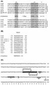
Alignment of promoter regions of genes transcribed by ςK RNA polymerase. (A) Sequences near the transcription start sites of genes transcribed by RNA polymerase containing ςK. Promoters for six genes transcribed in the absence of GerE and five genes whose transcription required GerE in addition to ςK are shown separately. Nucleotides in each promoter that match the consensus sequence are shown between the groups (m = C or A). The underlined nucleotides correspond to the transcription start point. (B) Alignment of nucleotide sequences of GerE-binding sites. The consensus sequence proposed by Zheng et al. (39) is shown at the top. Numbers refer to positions relative to the transcriptional start site. ∗, sequence from the opposite DNA strand. The bottom line shows an enlarged consensus sequence for GerE binding based on the sequence shown. R, purine; W, A or T; Y, pyrimidine. (C) Nucleotide sequence of the cotS promoter, showing putative −35 and −10 regions, the transcription start point (+1), and a ribosome-binding site (SD) (2). The boxed sequence is a putative GerE binding site in the cotS operon. References for the sequences of these promoters are as follows: sigK (spoIVCB), 20; cotA, 29; cotB and cotD, 39; cotC, 39 and 40; cotF, 9; gerE, 8; spoVK, 14; cotVXW, cotX, and cotYZ, 36; cotG, 26; and cotS, 2.

Similar articles

Cited by

References

    1. Abe A, Ogawa S, Kohno T, Watabe K. Purification of Bacillus subtilis spore coat protein by electrophoretic elution procedure and determination of NH2-terminal amino acid sequences. Microbiol Immunol. 1993;37:809–812. - PubMed
    1. Abe A, Koide H, Kohno T, Watabe K. A Bacillus subtilis spore coat polypeptide gene, cotS. Microbiology. 1995;141:1433–1442. - PubMed
    1. Adam D, Roels S, Beall B, Moran C P, Jr, Losick R. Subcellular localization of proteins involved in the assembly of the spore coat of Bacillus subtilis. Genes Dev. 1994;8:234–244. - PubMed
    1. Aronson A I, Fitz-James P C. Structure and morphogenesis of the bacterial spore coat. Bacteriol Rev. 1976;40:360–402. - PMC - PubMed
    1. Aronson A I, Song H-Y, Bourne N. Gene structure and precursor processing of a novel Bacillus subtilis spore coat protein. Mol Microbiol. 1989;3:437–444. - PubMed

MeSH terms

LinkOut - more resources