. 1998 Jun 1;141(5):1207-15.
doi: 10.1083/jcb.141.5.1207.

Differential subcellular localization of protein phosphatase-1 alpha, gamma1, and delta isoforms during both interphase and mitosis in mammalian cells

Affiliations

Differential subcellular localization of protein phosphatase-1 alpha, gamma1, and delta isoforms during both interphase and mitosis in mammalian cells

P R Andreassen et al. J Cell Biol. .

Abstract

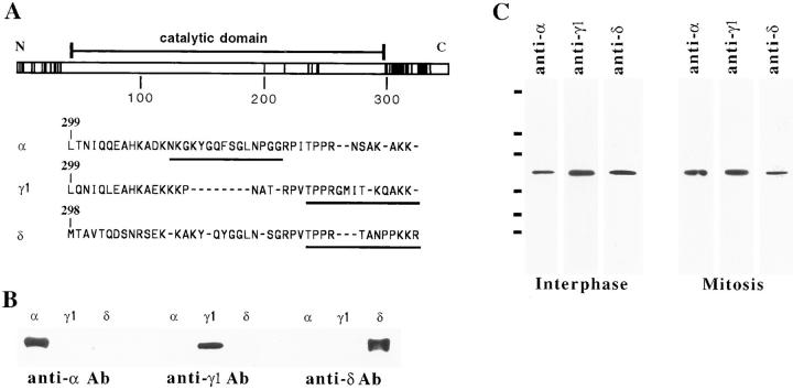
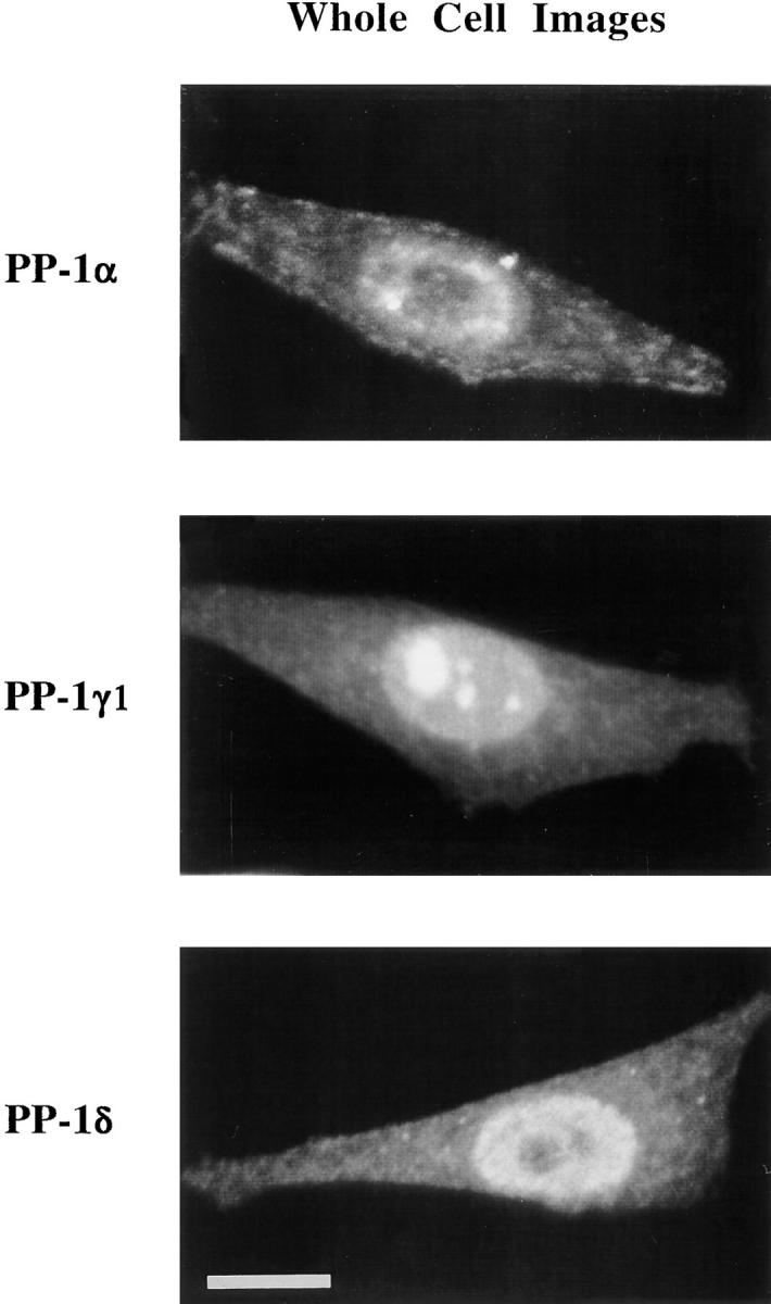
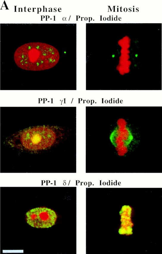
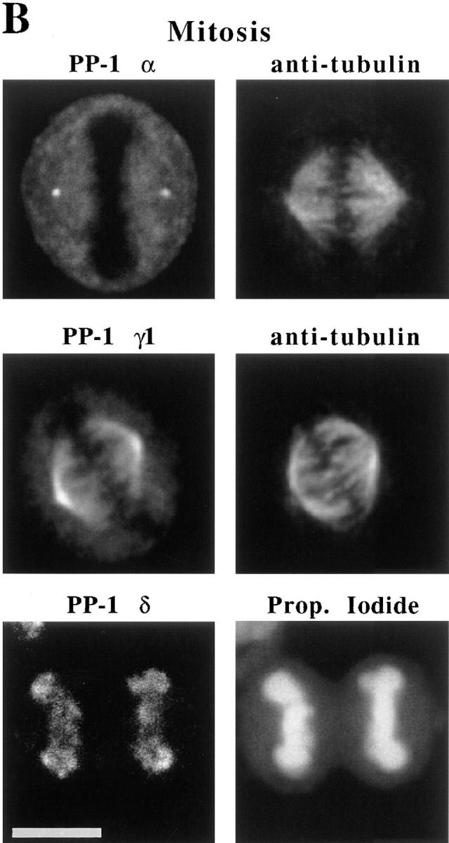
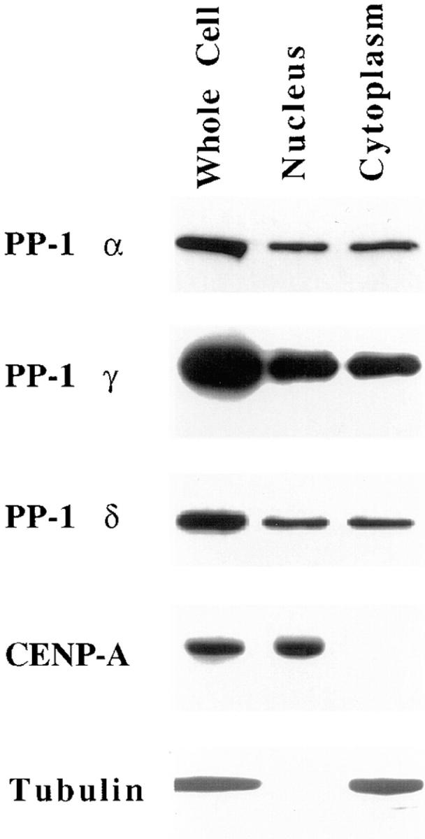
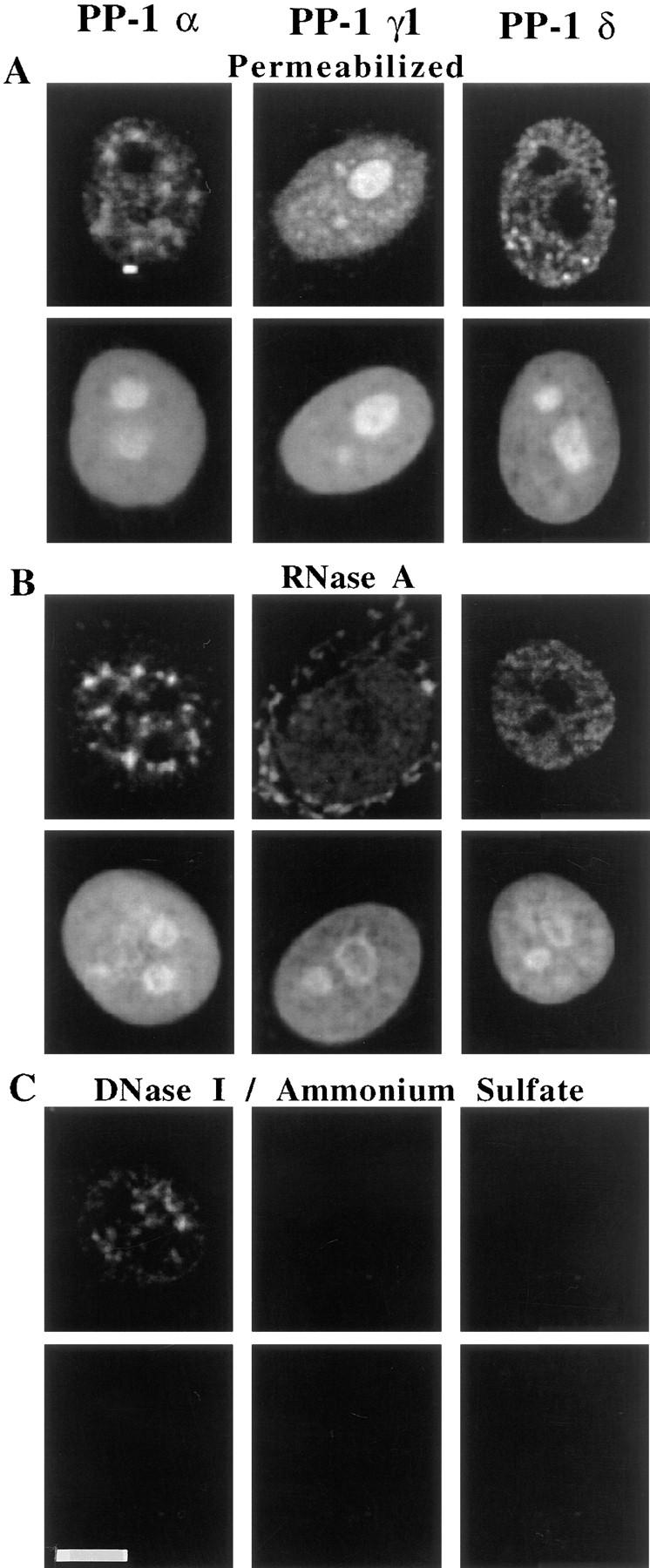
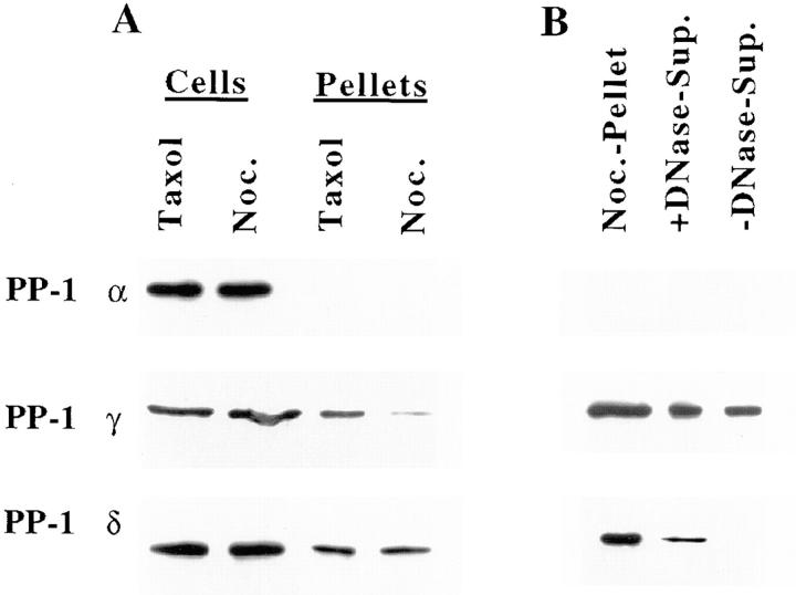
Protein phosphatase-1 (PP-1) is involved in the regulation of numerous metabolic processes in mammalian cells. The major isoforms of PP-1, alpha, gamma1, and delta, have nearly identical catalytic domains, but they vary in sequence at their extreme NH2 and COOH termini. With specific antibodies raised against the unique COOH-terminal sequence of each isoform, we find that the three PP-1 isoforms are each expressed in all mammalian cells tested, but that they localize within these cells in a strikingly distinct and characteristic manner. Each isoform is present both within the cytoplasm and in the nucleus during interphase. Within the nucleus, PP-1 alpha associates with the nuclear matrix, PP-1 gamma1 concentrates in nucleoli in association with RNA, and PP-1 delta localizes to nonnucleolar whole chromatin. During mitosis, PP-1 alpha is localized to the centrosome, PP-1 gamma1 is associated with microtubules of the mitotic spindle, and PP-1 delta strongly associates with chromosomes. We conclude that PP-1 isoforms are targeted to strikingly distinct and independent sites in the cell, permitting unique and independent roles for each of the isoforms in regulating discrete cellular processes.


Figures

Figure 1
Figure 1
Isoforms of PP-1 are most divergent at their COOH termini. (A) The sequences of human PP-1 α, γ1, and δ have been aligned and compared in this schematic representation. Each amino acid difference between any pair of PP-1 isoforms (Barker et al., 1994) is indicated by a solid vertical line. There is a modest amount of divergence at the NH2 terminus, but most divergence occurs proximal to the COOH terminus. There are few differences within the conserved catalytic domain (amino acids 42–98 in human PP-1 α; Peruski et al., 1993), indicated by a horizontal bar above the diagram. Single letter code sequences of PP-1 α, γ1, and δ at their divergent COOH termini are shown below the diagram. The peptide sequences used for the generation of isoform-specific antibodies are underlined. (B) Cross-blots of the PP-1 α, γ1, and δ antibodies against all three isoforms demonstrate that each antibody is isoform specific. (C) Antibodies generated against COOH-terminal peptides from PP-1 α, γ1, and δ each specifically recognize a 37-kD protein corresponding to PP-1 in blots of lysates from either interphase or mitotic HeLa cells. Bars at the left margin indicate size markers: 105, 82, 45, 33, 29, and 19 kD, respectively.
Figure 2
Figure 2
PP-1 α, γ1, and δ are each localized to both cytoplasm and nuclei in interphase HeLa cells. Images, representing whole cells, were generated from optical sections throughout the cell depth that were collected by confocal microscopy. A detailed comparison of isoform distributions in nuclei by optical sections of interphase cells is shown in Fig. 3 A. Bar, 10 μm.
Figure 3
Figure 3
PP-1 isoforms are differentially localized both during interphase and mitosis in HeLa cells. (A) Images for PP-1 α, γ1, and δ (green) merged with respective images of propidium iodide counterstain (red) are shown for cells at interphase and at mitotic metaphase. All primary antibodies were detected with FITC-conjugated secondary antibodies. Each isoform is present in the nucleus at interphase. PP-1 γ1 localizes to the nucleolus, whereas PP-1 α and δ distribute elsewhere in the nucleus. At mitotic metaphase, PP-1 α, γ1, and δ antigens are distributed to the centrosome, mitotic microtubules, and chromosomes, respectively. Nucleoli and chromosomes are strongly stained by propidium iodide at interphase and mitosis, respectively. Images are yellow where the green and red signals overlap. A centrosome adjacent to the nucleus of an interphase cell is labeled with PP-1 α–specific antibody. (B) Separated images show PP-1 isoforms are specifically associated either with the mitotic spindle, centrosomes, or chromosomes of HeLa cells. (Top) Double-label immunofluorescence images of PP-1 α (left) and antitubulin (right) in a metaphase cell demonstrate that PP-1 α is concentrated at the centrosome. (Middle) Double-label immunofluorescence images of PP-1 γ1 (left) and antitubulin (right) show that PP-1 γ1 is localized to spindle microtubules at metaphase. (Bottom) PP-1 δ (left) remains associated with chromosomes throughout mitosis, as shown here for a telophase cell. The propidium iodide counterstain (right) confirms that the cell is in telophase. Bars, 10 μm.
Figure 3
Figure 3
PP-1 isoforms are differentially localized both during interphase and mitosis in HeLa cells. (A) Images for PP-1 α, γ1, and δ (green) merged with respective images of propidium iodide counterstain (red) are shown for cells at interphase and at mitotic metaphase. All primary antibodies were detected with FITC-conjugated secondary antibodies. Each isoform is present in the nucleus at interphase. PP-1 γ1 localizes to the nucleolus, whereas PP-1 α and δ distribute elsewhere in the nucleus. At mitotic metaphase, PP-1 α, γ1, and δ antigens are distributed to the centrosome, mitotic microtubules, and chromosomes, respectively. Nucleoli and chromosomes are strongly stained by propidium iodide at interphase and mitosis, respectively. Images are yellow where the green and red signals overlap. A centrosome adjacent to the nucleus of an interphase cell is labeled with PP-1 α–specific antibody. (B) Separated images show PP-1 isoforms are specifically associated either with the mitotic spindle, centrosomes, or chromosomes of HeLa cells. (Top) Double-label immunofluorescence images of PP-1 α (left) and antitubulin (right) in a metaphase cell demonstrate that PP-1 α is concentrated at the centrosome. (Middle) Double-label immunofluorescence images of PP-1 γ1 (left) and antitubulin (right) show that PP-1 γ1 is localized to spindle microtubules at metaphase. (Bottom) PP-1 δ (left) remains associated with chromosomes throughout mitosis, as shown here for a telophase cell. The propidium iodide counterstain (right) confirms that the cell is in telophase. Bars, 10 μm.
Figure 4
Figure 4
Each PP-1 isoform is present in isolated nuclei. Immunoblots of Manca whole cell lysates, of isolated nuclei, and of the released cytoplasmic fraction are shown. All lanes were loaded on the basis of cell equivalence. The presence of each PP-1 isoform both in nuclei and cytoplasm is demonstrated by immunoblots probed for PP-1 α, γ1, and δ, respectively. Samples were also exposed to antitubulin antibodies to show the purity of isolated nuclei, and to CENP-A antiserum to show the purity of the cytoplasmic fractions.
Figure 5
Figure 5
Nuclear extraction demonstrates that the different PP-1 isoforms localize to distinct nuclear compartments. HeLa cells were fixed and prepared for immunofluorescence microscopy either (A) after permeabilization with Triton X-100, (B) permeabilization followed by RNA digestion, or (C) permeabilization followed by chromatin extraction through treatment with DNase I and then 0.25 M ammonium sulfate. PP-1 α, γ1, and δ are all present in nuclei after permeabilization. PP-1 γ1 is sensitive to RNA extraction, PP-1 δ is extracted with chromatin, and PP-1 α resists extraction through association with the nuclear matrix. Propidium iodide counterstain reveals both the extraction of nucleolar RNA by RNase treatment and the extraction of chromatin by the combination of DNase I and salt. Bar, 10 μm.
Figure 6
Figure 6
PP-1 isoforms distribute to different compartments in mitotic HeLa cells as assayed in immunoblots. PP-1 γ1, but not PP-1 α or δ, is enriched in microtubule fractions after cell lysis (A). Residual pellets from cells treated with taxol contain assembled microtubules, but pellets from cells treated with 1.0 μg/ml nocodazole (Noc.) are devoid of microtubules. PP-1 γ1 is preferentially associated with the residual cell pellet that contains microtubules after treatment with taxol. Whole cell lysates and residual cell pellets from cells treated with either taxol or nocodazole and loaded on the basis of equal cell number are shown. PP-1 δ is associated with chromosomes, as determined by release from the residual cell pellet after digestion with DNase I (B). Mitotic cells were collected by selective detachment, permeabilized, and incubated in digestion buffer either with (+) or without (−) DNase I, and supernatants (Sup.) were loaded onto gels. The residual cell pellet after lysis, loaded on the basis of equal cell number, is shown for reference. The majority of PP-1 δ is present in the cell supernatant from nocodazole-treated cells (see Fig. 6 A). Therefore, the blot of PP-1 δ shown here was exposed extensively to bring out the pellet fraction to examine its extractability by digestion with DNase I.

Similar articles

Cited by

References

    1. Alberts AS, Thorburn AM, Shenolikar S, Mumby MC, Feramisco JR. Regulation of cell cycle progression and nuclear affinity of the retinoblastoma protein by protein phosphatases. Proc Natl Acad Sci USA. 1993;90:388–392. - PMC - PubMed
    1. Alessi DR, Street AJ, Cohen P, Cohen PTW. Inhibitor-2 functions like a chaperone to fold three expressed isoforms of mammalian protein phosphatase-1 into a conformation with the specificity and regulatory properties of the native enzyme. Eur J Biochem. 1993;213:1055–1066. - PubMed
    1. Andreassen PR, Margolis RL. Microtubule dependency of p34cdc2inactivation and mitotic exit in mammalian cells. J Cell Biol. 1994;127:789–802. - PMC - PubMed
    1. Axton JM, Dombradi V, Cohen PTW, Glover DM. One of the protein phosphatase 1 isoenzymes in Drosophila is essential for mitosis. Cell. 1990;63:33–46. - PubMed
    1. Barker HM, Craig SP, Spurr NK, Cohen PTW. Sequence of human protein serine/threonine phosphatase 1 γ and localization of the gene (PPP1CC) encoding it to chromosome bands 12q24.1-q24.2. Biochim Biophys Acta. 1993;1178:228–233. - PubMed

Publication types