. 1998 Jun 1;141(5):1229-41.
doi: 10.1083/jcb.141.5.1229.

Defining the interactions between intermediate filaments and desmosomes

Affiliations

Defining the interactions between intermediate filaments and desmosomes

E A Smith et al. J Cell Biol. .

Abstract

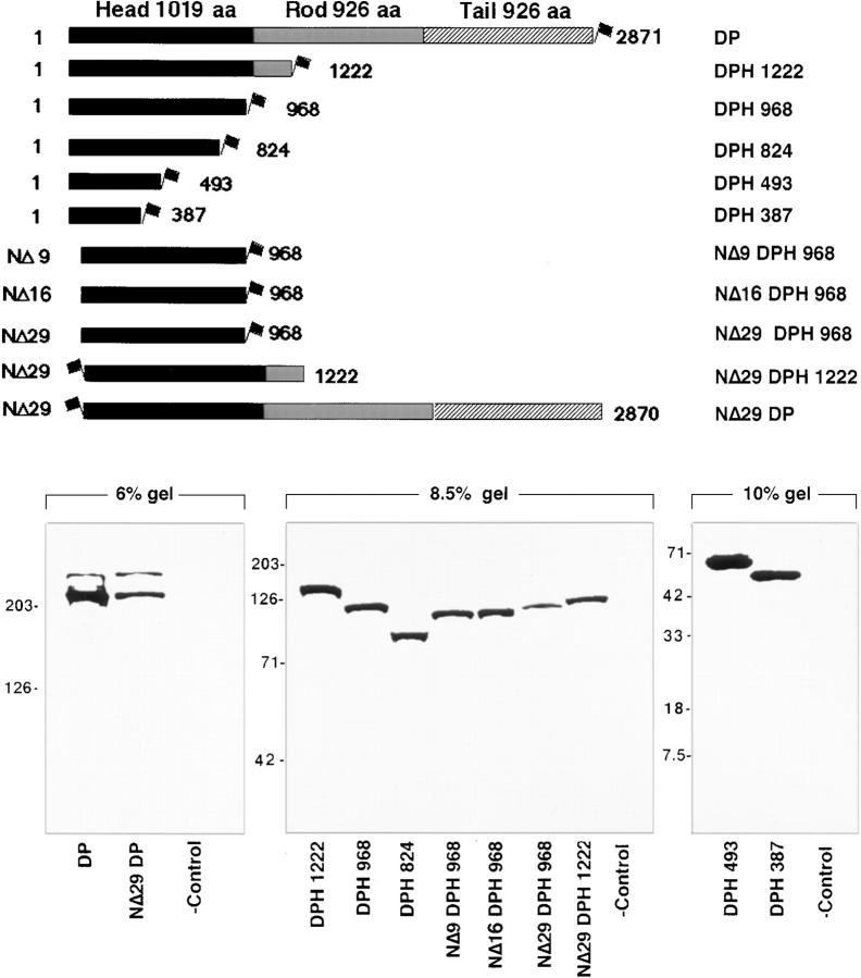
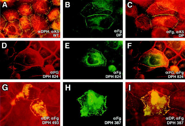
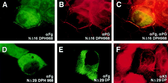
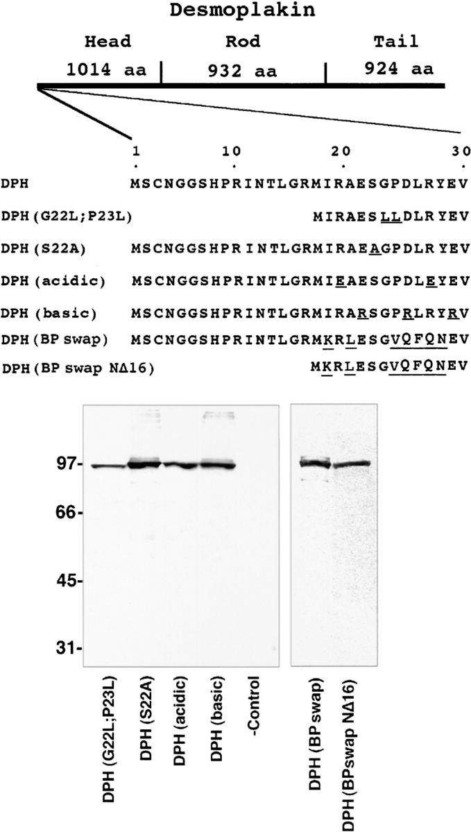
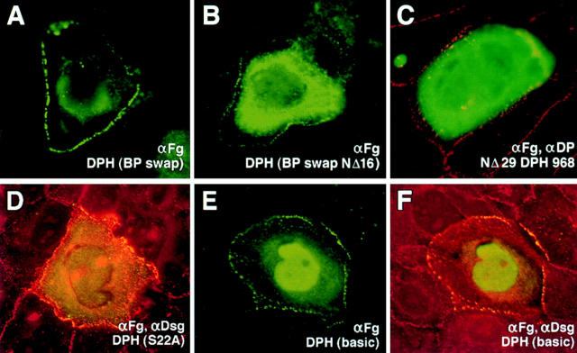
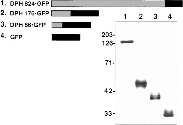
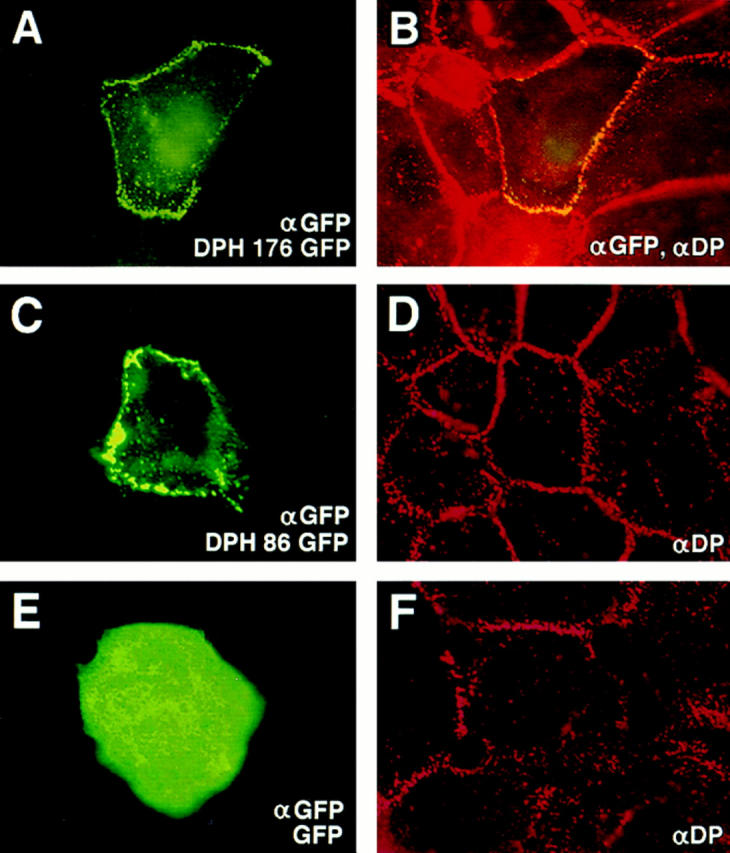
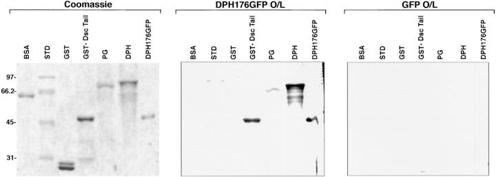
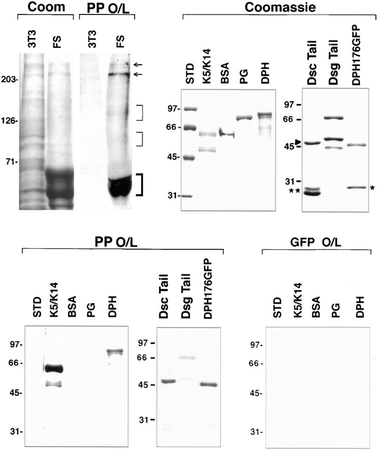
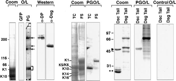
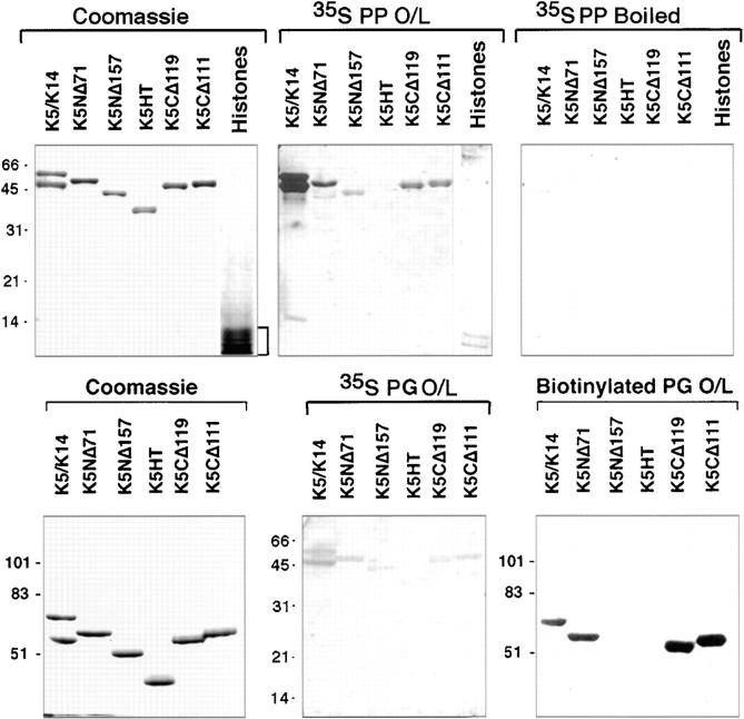
Desmoplakin (DP), plakoglobin (PG), and plakophilin 1 (PP1) are desmosomal components lacking a transmembrane domain, thus making them candidate linker proteins for connecting intermediate filaments and desmosomes. Using deletion and site-directed mutagenesis, we show that remarkably, removal of approximately 1% of DP's sequence obliterates its ability to associate with desmosomes. Conversely, when linked to a foreign protein, as few as 86 NH2-terminal DP residues are sufficient to target to desmosomes efficiently. In in vitro overlay assays, the DP head specifically associates with itself and with desmocollin 1a (Dsc1a). In similar overlay assays, PP1 binds to DP and Dsc1a, and to a lesser extent, desmoglein 1 (Dsg1), while PG binds to Dsg1 and more weakly to Dsc1a and DP. Interestingly, like DP, PG and PP1 associate with epidermal keratins, although PG is considerably weaker in its ability to do so. As judged by overlay assays, the amino terminal head domain of type II keratins appears to have a special importance in establishing these connections. Taken together, our findings provide new insights into the complexities of the links between desmosomes and intermediate filaments (IFs). Our results suggest a model whereby at desmosome sites within dividing epidermal cells, DP and PG anchor to desmosomal cadherins and to each other, forming an ordered array of nontransmembrane proteins that then bind to keratin IFs. As epidermal cells differentiate, PP1 is added as a molecular reinforcement to the plaque, enhancing anchorage to IFs and accounting at least partially for the increase in numbers and stability of desmosomes in suprabasal cells.


Figures

Figure 1
Figure 1
Desmoplakin deletion mutants and their expression in cultured epithelial cells. (Top) Shown are diagrams of truncated mutant desmoplakin proteins whose expression was genetically engineered to be under the control of the SV40 major early promoter and enhancer. In all cases, the FLAG epitope tag was placed in-frame (depicted by the flag). The full-length DP transgene was engineered from part DP cDNA and part DP gene, such that both DPI and DPII would be expressed. The coiled coil rod and flanking globular head and tail segments are demarcated according to the Coils Version 2.1 secondary structure prediction program (www site, ISREC, Switzerland). Numbers shown are in amino acid residues; nomenclature is at right. (Bottom) Western blot analyses of protein extracts from COS epithelial cells transfected with the constructs indicated above and harvested 48 h after transfection. Proteins were resolved by electrophoresis through SDS polyacrylamide gels (percentages noted), transferred to nitrocellulose paper by electroblotting, and then visualized by binding to anti-FLAG. Antibody binding was developed using biotinylated secondary antibodies as outlined by Kouklis et al. (1994). (-control) Extracts from mock-transfected cells. Migration of size standards are indicated at left, in kD. Since some transfections were conducted at different times and since variations existed in plasmid preparations and transfection efficiencies, loadings were done to reflect similar transgene protein levels, rather than total protein levels.
Figure 2
Figure 2
Localization of full-length and COOH-terminally truncated desmoplakins to desmosomes. Human epidermal keratinocytes (SCC-13) were transfected with expression vectors encoding proteins denoted at the lower right of each frame. After transfection, cells were fixed and labeled with antibodies indicated, followed by the appropriate fluorescently conjugated secondary antibodies. Representative cells shown were visualized through a Zeiss Axiophot immunofluorescence microscope with a 100×or 63× objective. Punctate staining at cell borders is typical of desmosomal staining; filamentous staining is typical of keratin network staining. WT, wild-type.
Figure 3
Figure 3
Localization of NH2-terminally truncated desmoplakins to desmosomes. Human epidermal keratinocytes were transfected with expression vectors encoding FLAG epitope–tagged desmoplakin proteins. After transfection, cells were fixed and stained with the indicated antibodies. Representative cells were photographed using a 100× objective. Note loss of punctate staining and appearance of diffuse cytoplasmic staining in transfected cells in D; note loss of punctate staining and appearance of filamentous staining in transfected cell in E and F.
Figure 4
Figure 4
Construction and expression of mutants within N16-N29 of the desmoplakin head domain. (Top) Diagram depicting the first 29 amino acid residues of the DPH and the various constructs engineered in which residues were mutated within this sequence. The most dramatic change was the swapping of the N16-N29 residues within the equivalent sequence of BPAG1e to create DPH (BPswap), which was engineered in the context of the full-length head domain, and in the context of DPH NΔ16. (Bottom)Immunoblot anlaysis of extacts isolated from cells 48 h after transfection with vectors encoding these proteins. (-control) Extract from mock-transfected cells. Molecular mass standards in kD are indicated at left.
Figure 5
Figure 5
Changes in the localization of the desmoplakin head domain upon mutation within the first 29 NH2-terminal residues of DPH. Keratinocytes were transfected with the vectors encoding mutant DPH proteins. After transfection, cells were fixed and stained with antibodies as indicated. Note progressive loss of cell border staining going from DPH (BPswap) in A to DPH(BPswapNΔ16) in B to NΔ29DPH in C; note addition of nuclear staining in E and F.
Figure 6
Figure 6
Construction and expression of DPH fusion proteins. (Top) Diagram indicating fusion proteins generated from portions of DPH (grey) linked to GFP (black bars). (Bottom) Cells were transfected with expression vectors encoding the DPH-GFP fusion proteins. After transfection, extracts were isolated and subjected to electrophoresis through 10% SDS polyacrylamide gels, electroblotted onto nitrocellulose paper, probed with anti-GFP and subjected to chemiluminescence. Molecular mass standards in kD at left.
Figure 7
Figure 7
Localization of desmoplakin fusion proteins to desmosomes. Keratinocytes were transfected with expression vectors encoding desmoplakin fusion proteins. After transfection, cells were fixed and stained with the indicated antibodies (lower right). The αDP used was to the tail segment of DP, not present in the fusion protein. Representative cells for each construct were photographed under an 100× objective, and frames are displayed here in pairs, with transgene product staining at left and double-staining at right. Note that cells expressing DPH 176 GFP or DPH 86 GFP displayed predominantly punctate αGFP/αDP staining at the periphery, typical of desmosomal localization; in contrast, cells expressing GFP exhibited cytoplasmic staining with little or no membrane staining.
Figure 8
Figure 8
Evidence for direct interaction between 176 residues of DPH and the cytoplasmic domain of Dsc1a and the DPH itself. Recombinant proteins were engineered as described in Materials and Methods, and resolved in triplicate by SDS PAGE. One gel was stained with Coomassie Blue to visualize the proteins, and the others were transferred to nitrocellulose membrane and then subjected to overlay assays using equal amounts of [S35]methionine-labeled DPH176GFP (test) and GFP (control), respectively. Note binding of DPH176GFP to the Dsc1a tail, DPH, and DPH176GFP, under conditions where radiolabeled GFP showed no binding. Molecular mass standards in kilodaltons at left.
Figure 9
Figure 9
Evidence for direct interaction between plakophilin 1 and desmosomal proteins. (Top, from left to right) Proteins from the Triton X-100 insoluble fraction of mouse 3T3 fibroblasts or human foreskin were resolved by electrophoresis through a 6% SDS polyacrylamide gel, and were either stained with Coomassie Blue to visualize proteins (Coom) or subjected to overlay assays (O/L) using S35-methionine–labeled plakophilin 1 (PP). Note binding of PP to bands of the mobility of desmoplakins I and II (arrows), and to keratins (bold bracket). Note also faint binding to two additional groups of skin bands (thin brackets), which were of the mobility of the desmogleins and desmocollins. Interactions between PP1 and desmosomal components were verified using recombinant proteins. Coomassie Blue–stained gels of bacterially expressed and purified recombinant proteins are shown, along with overlay assays using equal amounts of S35-labeled PP1 (test) and GFP (control). Double asterisk and single asterisk denote GST and GFP, respectively, added to the lanes containing DscTail and DPH176GFP as internal controls. Dsctail, GST-Dsc1a-tail fusion protein; Dsgtail, Dsg1-tail; Std, molecular mass standards (sizes denoted in kD at left).
Figure 10
Figure 10
Evidence for direct interaction between plakoglobin and a number of desmosomal proteins. (Top, from left to right) Proteins from the Triton X-100–insoluble fraction of human foreskin were resolved by electrophoresis through a 6% SDS polyacrylamide gel and either stained with Coomassie Blue to visualize proteins (Coom) or subjected to overlay assays (O/L) using equivalent amounts of [S35]methionine- labeled GFP (control) or plakoglobin (PG, test). Note clear binding of PG to bands of the mobility of desmoplakins (top arrow), desmogleins (bottom two arrows), and to keratins (bracket). Western blot analysis confirmed the presence of desmoplakins and desmogleins in this extract. Binding of PG to the keratins was confirmed using extracts from human epidermal cultures (TC) and from foreskin (FS), depicting predominantly K5/6 interactions in TC extracts and K1 interactions in FS extracts. Interactions between PG and the desmosomal cadherins were further examined using recombinant proteins. Coomassie Blue–stained gels of bacterially expressed and purified GST-Dsc1a-tail (Dsc Tail) fusion protein and recombinant Dsg1-tail (Dsg Tail) are shown, along with overlay assays using equal amounts of S35-labeled PG. Double asterisk denotes GST, added to the lane containing DscTail as an internal control. Molecular mass standards are shown in kD at left.
Figure 11
Figure 11
Comparison of the interactions among PP1, PG, and keratins. Interactions between keratins and plakophilin/plakoglobin were examined in greater detail using recombinant keratins and overlay assays with equal amounts of in vitro–translated S35-labeled PP1 and S35-labeled PG proteins that we determined to have comparable specific activities. Overlays were also performed with biotinylated recombinant PG. As a control for nonspecific charge interactions, we included a mixture of bovine histones (Sigma Chemical Co., St. Louis, MO) and a control, whereby the probe was boiled before hybridization. Recombinant K5 mutant proteins were as described previously (Wilson et al., 1992; Kouklis et al., 1994). Note that under equivalent conditions, the interactions with keratins were significantly stronger for PP1 than PG, and that the more sensitive biotinylated PG probe was needed to analyze PG-IF interactions. Note also that only those K5 proteins with an intact amino terminal domain hybridized with the armadillo probes.

References

    1. Barth AI, Nathke IS, Nelson WJ. Cadherins, catenins and APC protein: interplay between cytoskeletal complexes and signaling pathways. Curr Opin Cell Biol. 1997;9:683–690. - PubMed
    1. Bierkamp C, McLaughlin KJ, Schwarz H, Huber O, Kemler R. Embryonic heart and skin defects in mice lacking plakoglobin. Dev Biol. 1996;180:780–785. - PubMed
    1. Bornslaeger EA, Corcoran CM, Stappenbeck TS, Green KJ. Breaking the connection: displacement of the desmosomal plaque protein desmoplakin from cell-cell interfaces disrupts anchorage of intermediate filament bundles and alters intercellular junction assembly. J Cell Biol. 1996;134:985–1001. - PMC - PubMed
    1. Buxton RS, Cowin P, Franke WW, Garrod DR, Green KJ, King IA, Koch PJ, Magee AI, Rees DA, Stanley JR, Steinberg MS. Nomenclature of the desmosomal cadherins. J Cell Biol. 1993;121:481–483. - PMC - PubMed
    1. Candi E, Tarcsa E, Digiovanna JJ, Compton JG, Elias PM, Marekov LN, Steinert PM. A highly conserved lysine residue on the head domain of type II keratins is essential for the attachment of keratin intermediate filaments to the cornified cell envelope through isopeptide crosslinking by transglutaminases. Proc Natl Acad Sci USA. 1998;95:2067–2072. - PMC - PubMed

Publication types