. 1998 Jun 1;141(5):1277-86.
doi: 10.1083/jcb.141.5.1277.

Lumican regulates collagen fibril assembly: skin fragility and corneal opacity in the absence of lumican

Affiliations

Lumican regulates collagen fibril assembly: skin fragility and corneal opacity in the absence of lumican

S Chakravarti et al. J Cell Biol. .

Abstract

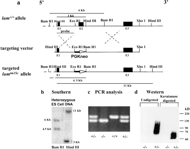
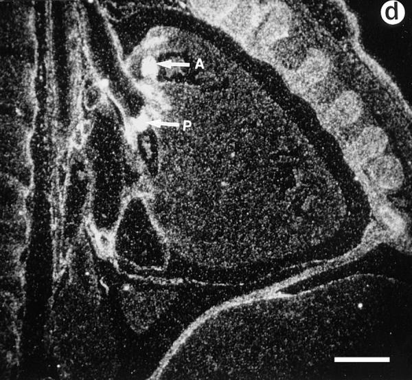
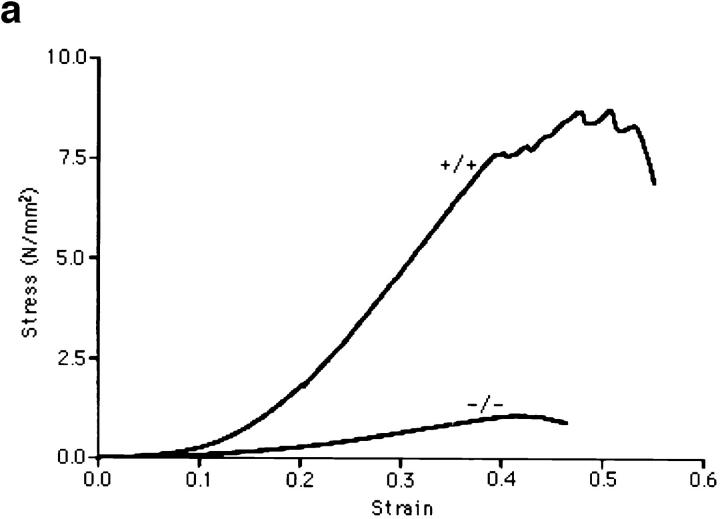
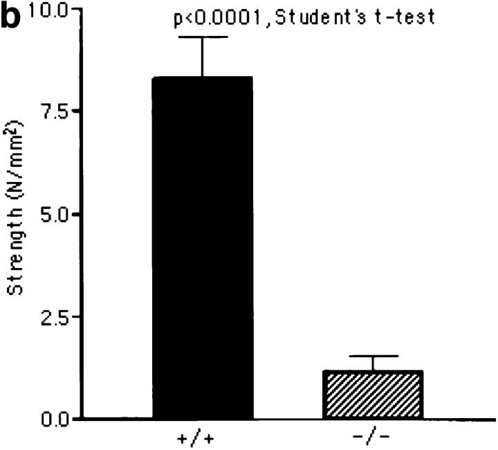
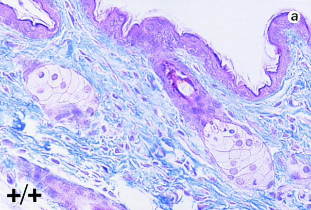
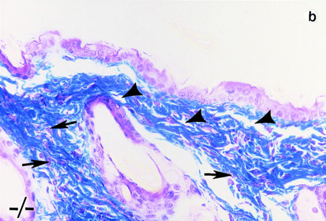
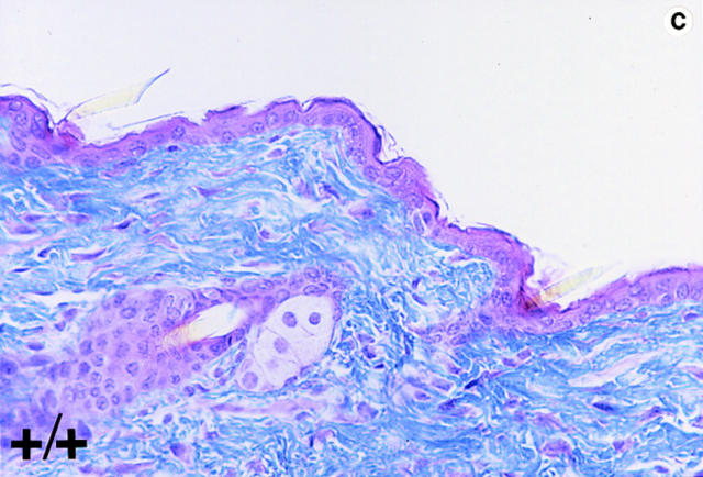
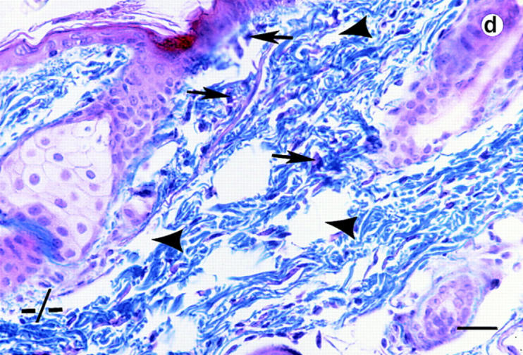
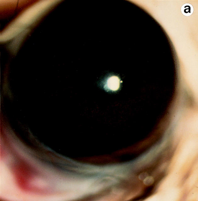
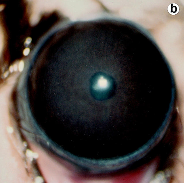
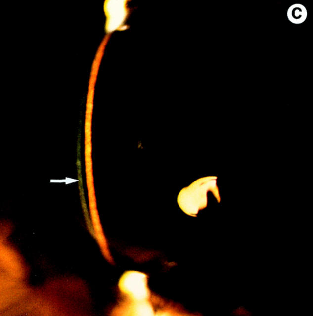
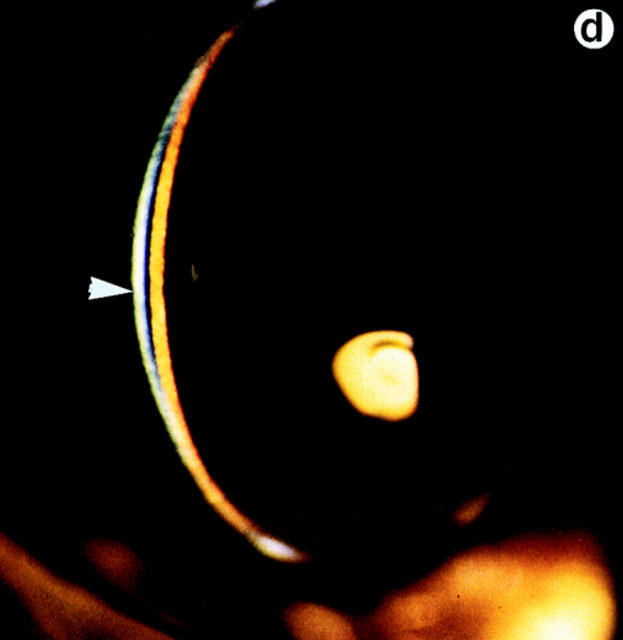
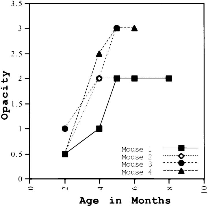
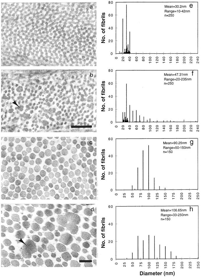
Lumican, a prototypic leucine-rich proteoglycan with keratan sulfate side chains, is a major component of the cornea, dermal, and muscle connective tissues. Mice homozygous for a null mutation in lumican display skin laxity and fragility resembling certain types of Ehlers-Danlos syndrome. In addition, the mutant mice develop bilateral corneal opacification. The underlying connective tissue defect in the homozygous mutants is deregulated growth of collagen fibrils with a significant proportion of abnormally thick collagen fibrils in the skin and cornea as indicated by transmission electron microscopy. A highly organized and regularly spaced collagen fibril matrix typical of the normal cornea is also missing in these mutant mice. This study establishes a crucial role for lumican in the regulation of collagen assembly into fibrils in various connective tissues. Most importantly, these results provide a definitive link between a necessity for lumican in the development of a highly organized collagenous matrix and corneal transparency.


Figures

Figure 2
Figure 2
Gene targeting at the lum locus. Exons are marked as E1, E2, and E3. (a) Diagram of the wild-type lum + allele, targeting vector, and targeted lumtm1Sc allele. Correct targeting should replace the 3-kb EcoRI–BamHI segment containing exon 2 (E2) with the 1.33-kb pGKneo cassette for positive selection. (b) Diagnostic Southern hybridization on ES clone DNA. Presence of a 4.5-kb BamHI and an 11-kb HindIII fragment indicates one correctly targeted allele. (c) PCR analysis of offsprings from heterozygous matings. (d) Western blot analysis on total protein extracted from whole eyes of lumtm1Sc/ lumtm1Sc (−/−) and lum +/lumtm1Sc (+/−) mice. Undigested extract from heterozygous mice show a broad band at 65–90 kD, indicating the presence of lumican. Lack of a sharp band is due to heterogenous KS modification of lumican. After keratanase treatment and removal of most of the KS a sharper and faster migrating band results, a characteristic feature of KS proteoglycans. The lumtm1Sc/lumtm1Sc tissue lacks any immunoreactive material confirming the absence of any lumican gene product.
Figure 1
Figure 1
Lumican expression during mouse embryonic development. In situ hybridization analysis. (a) Sagittal section of E.9.5. Lumican is expressed at high levels in head (arrow) and lateral mesenchyme (arrowhead). (b) Transverse section of E13.5 embryo. At E13.5 lumican expression is focused in the subepithelial dermis of eyelid area (asterisk), cornea (arrowhead), with lesser amounts in the sclera (arrow). The lens and retina do not express lumican. (c) Section through E15.5 stage dorsal skin. Top panel is a bright field view of a toluidine blue–stained section, and bottom panel is a dark field view of same section showing silver grain deposits. Note that darkly stained epidermal layer (Ep) does not contain lumican mRNA. Dermal layer (Dm) displays a strong signal for lumican transcript. No lumican mRNA in central canal in sacro-coccygeal region of spinal cord (Sp). (d) Parasagittal section through E15.5 embryo showing heart pulmonary (P) and aortic valve (A) leaflets with strong signal for lumican transcript. Bars, 100 μm.
Figure 1
Figure 1
Lumican expression during mouse embryonic development. In situ hybridization analysis. (a) Sagittal section of E.9.5. Lumican is expressed at high levels in head (arrow) and lateral mesenchyme (arrowhead). (b) Transverse section of E13.5 embryo. At E13.5 lumican expression is focused in the subepithelial dermis of eyelid area (asterisk), cornea (arrowhead), with lesser amounts in the sclera (arrow). The lens and retina do not express lumican. (c) Section through E15.5 stage dorsal skin. Top panel is a bright field view of a toluidine blue–stained section, and bottom panel is a dark field view of same section showing silver grain deposits. Note that darkly stained epidermal layer (Ep) does not contain lumican mRNA. Dermal layer (Dm) displays a strong signal for lumican transcript. No lumican mRNA in central canal in sacro-coccygeal region of spinal cord (Sp). (d) Parasagittal section through E15.5 embryo showing heart pulmonary (P) and aortic valve (A) leaflets with strong signal for lumican transcript. Bars, 100 μm.
Figure 3
Figure 3
Skin tensile stress-strain measurements. (a) Representative tensile stress-strain curves for skin samples from wild type (+/+) and lumtm1Sc homozygous null mutants (−/−), n = 5 for each genotype. Tension tests were performed in duplicate on freshly isolated skin samples with a 10 × 20-mm gauge region from the dorsal surface of each animal. Samples were loaded to failure in tension at a constant rate of 1 mm/s using an Instron servohydraulic materials test machine. Stress in N/mm2 was plotted versus the strain. Note the significant reduction in both the modulus and the stress to failure in the −/− animals. (b) Tensile strength in wild type (+/+) and lumtm1Sc homozygous null mutants (−/−). The average tensile strength or maximum stress in the (−/−) skin was 86% lower than the wild-type (+/+) skin.
Figure 3
Figure 3
Skin tensile stress-strain measurements. (a) Representative tensile stress-strain curves for skin samples from wild type (+/+) and lumtm1Sc homozygous null mutants (−/−), n = 5 for each genotype. Tension tests were performed in duplicate on freshly isolated skin samples with a 10 × 20-mm gauge region from the dorsal surface of each animal. Samples were loaded to failure in tension at a constant rate of 1 mm/s using an Instron servohydraulic materials test machine. Stress in N/mm2 was plotted versus the strain. Note the significant reduction in both the modulus and the stress to failure in the −/− animals. (b) Tensile strength in wild type (+/+) and lumtm1Sc homozygous null mutants (−/−). The average tensile strength or maximum stress in the (−/−) skin was 86% lower than the wild-type (+/+) skin.
Figure 4
Figure 4
Histological analyses on skin alterations. Sections through skin from dorsal (a and b) and ventral (c and d) surfaces. Wild-type animals (+/+) shown in a and c and lumican null mutants (−/−) shown in b and d. The sections were trichrome stained in which the connective tissue, collagens primarily stain blue. Note disoriented fibroblasts (arrows) and open spaces (arrowheads) in the dermis of b and d. Bar, 50 μm.
Figure 4
Figure 4
Histological analyses on skin alterations. Sections through skin from dorsal (a and b) and ventral (c and d) surfaces. Wild-type animals (+/+) shown in a and c and lumican null mutants (−/−) shown in b and d. The sections were trichrome stained in which the connective tissue, collagens primarily stain blue. Note disoriented fibroblasts (arrows) and open spaces (arrowheads) in the dermis of b and d. Bar, 50 μm.
Figure 5
Figure 5
Eye examination for corneal clarity. Full view photobiomicroscopy of wild type (a) and lumtm1Sc homozygous mutants (b) eyes. Wild-type mice show clear corneas through which iris details can be seen (a). A continuous cloudiness with a clear ringlike peripheral zone is noticeable in the null mutant (b). Slit-lamp photobiomicrography of wild type (c) and lumtm1Sc homozygous mutant (d). Note clear beam with normal granularity in c and enhanced brightness because of increased light scattering and extensive granular material in d.
Figure 5
Figure 5
Eye examination for corneal clarity. Full view photobiomicroscopy of wild type (a) and lumtm1Sc homozygous mutants (b) eyes. Wild-type mice show clear corneas through which iris details can be seen (a). A continuous cloudiness with a clear ringlike peripheral zone is noticeable in the null mutant (b). Slit-lamp photobiomicrography of wild type (c) and lumtm1Sc homozygous mutant (d). Note clear beam with normal granularity in c and enhanced brightness because of increased light scattering and extensive granular material in d.
Figure 6
Figure 6
Changes in corneal opacification over time. Out of 14 lumtm1Sc homozygous mutants that were given follow-up eye exams over 7–8 mo, progression in opacity is shown for four mutants here. All 14 animals showed increase in opacity over time.
Figure 7
Figure 7
Transmission electron micrography of cornea (a and b) and tail dermis (c and d) collagen preparations (17). (a and c) Homozygous wild type, and (b and d) Lumtm1Sc homozygous. Note larger abnormal shaped fibrils in cross section (arrowhead) in b and d. Fibril diameter frequency (e–h). Homozygous wild-type cornea and tail collagen (e and g) and lumtm1Sc homozygous cornea and tail collagen (f and h). Bar, 0.2 μm.

References

    1. Birk DE, Fitch JM, Babiarz JP, Doane KJ, Linsenmayer TE. Collagen fibrillogenesis in vitro: interactions of types I and V collagen regulates fibril diameter. J Cell Sci. 1990;95:649–657. - PubMed
    1. Blochberger TC, Vergnes J-P, Hempel J, Hassell JR. cDNA to chick lumican (corneal keratan sulfate proteoglycan) reveals homology to the small interstitial proteoglycan gene family and expression in muscle and intestine. J Biol Chem. 1992;267:347–352. - PubMed
    1. Burch GH, Gong Y, Liu W, Dettman RW, Curry CJ, Smith L, Miller WL, Bristow J. Tenascin-X deficiency is associated with Ehlers-Danlos syndrome. Nat Genet. 1997;17:104–108. - PubMed
    1. Byers PH, Holbrook KA, Hall JG, Bornstein P, Chandler JW. A new variety of spondyoepiphyseal dysplasia characterized by punctate corneal dystrophy and abnormal collagen fibrils. Hum Genet. 1978;40:157–169. - PubMed
    1. Casey, T.A., and K.W. Sharif. 1991. A Color Atlas of Corneal Dystrophies and Degenerations. Wolfe Publishing Limited, London. 21–51.

Publication types