. 1998 Jun 1;187(11):1767-78.
doi: 10.1084/jem.187.11.1767.

Early regeneration of thymic progenitors in rhesus macaques infected with simian immunodeficiency virus

Affiliations

Early regeneration of thymic progenitors in rhesus macaques infected with simian immunodeficiency virus

J J Wykrzykowska et al. J Exp Med. .

Abstract

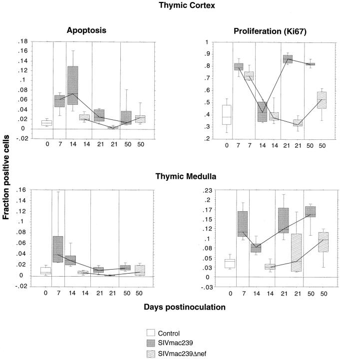
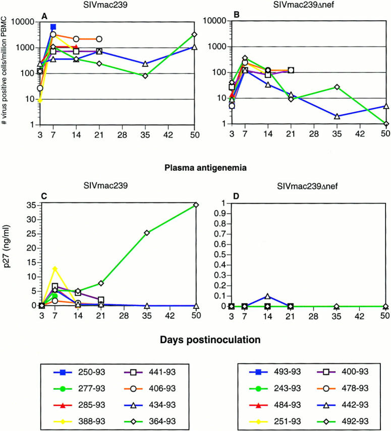
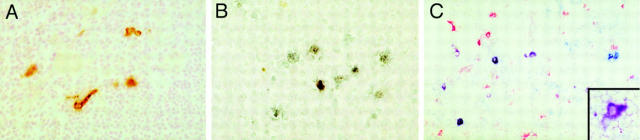
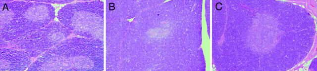
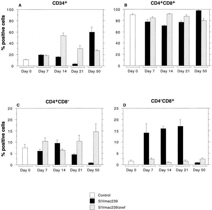
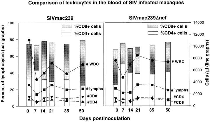
The thymus plays a critical role in the maturation and production of T lymphocytes and is a target of infection by human immunodeficiency virus (HIV) and the related simian immunodeficiency virus (SIV). Using the SIV/macaque model of AIDS, we examined the early effects of SIV on the thymus. We found that thymic infection by SIV resulted in increased apoptosis 7-14 d after infection, followed by depletion of thymocyte progenitors by day 21. A marked rebound in thymocyte progenitors occurred by day 50 and was accompanied by increased levels of cell proliferation in the thymus. Our results demonstrate a marked increase in thymic progenitor activity very early in the course of SIV infection, long before marked declines in peripheral CD4(+) T cell counts.


Figures

Figure 4
Figure 4
Apoptosis and cellular proliferation in the thymus during SIV infection. Alterations in cell proliferation and apoptosis in the thymic cortex and medulla were examined by morphometry in sections of thymus from animals infected with pathogenic SIVmac239 or nonpathogenic SIVmac239Δnef and compared with four uninfected control animals. Results are illustrated as box-plots and median traces. The box plots are based on 10 observations (20 for the controls), 5 from each animal. Each box-plot represents pooled data from two animals infected with the same virus or four uninfected age-matched controls. To examine cell proliferation, the fraction of cells positive for the Ki67 nuclear proliferation antigen were measured separately in the cortex and medulla using a Quantimet 570c image analyzer. The fraction of cells undergoing apoptosis was similarly determined after sections were subjected to in situ end labeling using the Apotag kit. The most dramatic changes occurred in the thymic cortex of animals infected with SIVmac239, where there was an increase in apoptosis at 14 dpi followed by an increase in proliferation at 21–50 dpi. Note that the scale of the Y axis for proliferation in the thymic cortex goes to 1, indicating that at 21 and 50 dpi nearly all of the cells in the cortex were proliferating. With the exception of an early increase in proliferation in the cortex, animals infected with SIVmac239Δnef showed no significant differences from the uninfected control group.
Figure 1
Figure 1
Quantitative virus isolation. Cell associated viral loads (A and B) and plasma antigenemia (C and D) in animals inoculated with pathogenic SIVmac239 (A and C) or nonpathogenic SIVmac239Δnef (B and D). Peak viral loads were seen at 7 dpi. Viral loads in animals infected with SIVmac239 were maintained at high levels, whereas viral loads in animals infected with SIVmac239Δnef declined to very low levels, by 50 dpi. Plasma antigenemia was transient in all animals except for 364–93, which was infected with SIVmac239. This animal demonstrated a typical “rapid progressor” profile with increasing plasma antigenemia and absence of a detectable humoral immune response.
Figure 2
Figure 2
Localization of SIVmac239 in the thymus. Immunohistochemistry for SIV gp120 (A) and in situ hybridization for SIV nucleic acid (B) revealed many infected cells in the thymus, particularly in the medulla. The most numerous infected cells were small round to oval cells, consistent with lymphocytes. In addition, as shown in A, scattered cells with distinct processes were also found to be infected in the medulla. To better determine the immunophenotype of the cells with dendritic morphology, double-label immunohistochemistry was performed (C), combining detection of SIVgp120 (blue) with a marker of monocyte/macrophages (CD68, red). In addition to individual cells positive for CD68 only (red) or SIVgp120 only (blue), several cells are double labeled in shades of purple due to mixing of red and blue chromogens. These purple cells are SIV-infected cells of monocyte/macrophage lineage. At higher magnification (inset), most of the infected cells with dendritic morphology were purple, indicating that they are of monocyte/macrophage lineage. Original magnification: A, ×80; B, ×100; C, ×75; inset, ×150.
Figure 3
Figure 3
Morphologic alterations of the thymus. Hematoxylin and eosin stained sections of thymus from macaques infected with SIVmac239 at 14 (A) and 50 (B) dpi and from an age-matched, uninfected macaque (C). The photomicrographs were taken at the same original magnification (×33). As compared with the uninfected control (C), moderate thinning of the cortex is visible at 14 dpi (A), whereas at 50 dpi (B) the thymic cortex is at least as thick as in the uninfected control.
Figure 5
Figure 5
T lymphocyte progenitors in the thymus. Flow cytometric analysis of thymocyte progenitors was performed on single cell suspensions of thymus collected at the time of euthanasia. Three-color flow cytometry was performed with antibodies to surface CD3, CD4, CD8, and CD34. The figure shows the proportion of positive cells for controls and for animals infected with SIVmac239 or SIVmac239Δnef at each time point. Bar heights indicate mean percentage of positive cells and whiskers above bars show one SEM. Animals infected with SIVmac239 had a decrease in both CD34+ and CD4+ CD8+ populations through d 21 followed by a dramatic rebound by 50 dpi. Marked changes in CD4 and CD8 single-positive cells were also seen in the thymus in animals infected with SIVmac239 including a striking increase in CD4CD8+ cells through d 21 and a marked decrease in the relative percentage of both CD4 and CD8 single-positive cells by d 50. The latter is likely due to the relative increase in the percentage of CD34+ and CD4+CD8+ T cell progenitors. Animals infected with SIVmac239Δnef showed few significant changes with the exception of an increase in CD34+ cells at 14 dpi.
Figure 6
Figure 6
Flow cytometric analysis of peripheral blood leukocytes in animals before and after infection with SIVmac239 or SIVmac239Δnef. Animals inoculated with SIVmac239 show a mild decline in CD4/CD8 ratio associated with a minimal decrease in CD4+ T cells and a small increase in CD8 cells. No significant alterations in CD4+ or CD8+ T cell numbers, percentages, or CD4/CD8 ratios were observed in animals inoculated with SIVmac239Δnef.

Similar articles

Cited by

References

    1. Spits H, Lanier LL, Phillips JH. Development of human T and natural killer cells. Blood. 1995;85:2654–2670. - PubMed
    1. Haynes BF, Denning SM, Le PT, Singer KH. Human intrathymic T cell differentiation. Semin Immunol. 1990;2:67–77. - PubMed
    1. Schuurman HJ, Krone WJA, Broekhuizen R, Baarlen JV, vanVeen P, Goldstein AL, Huber J, Goudsmit J. The thymus in acquired immune deficiency syndrome: comparison with other types of immunodeficiency disease and presence of components of human immunodeficiency virus type 1. Am J Pathol. 1989;134:1329–1334. - PMC - PubMed
    1. Burke AP, Anderson D, Benson W, Turnicky R, Mannan P, Liang YH, Smialek J, Virmani R. Localization of human immunodeficiency virus 1 RNA in thymic tissues from asymptomatic drug addicts. Arch Pathol Lab Med. 1995;119:36–41. - PubMed
    1. Schnittman SM, Denning SM, Greenhouse JJ, Justement JS, Baseler M, Kurtzberg J, Haynes BF, Fauci AS. Evidence for susceptibility of intrathymic T-cell precursors and their progeny carrying T-cell antigen receptor phenotypes TCRαβ+ and TCRγδ + to human immunodeficiency virus infection: a mechanism for CD4+(T4) lymphocyte depletion. Proc Natl Acad Sci USA. 1990;87:7727–7731. - PMC - PubMed

Publication types