. 1998 Jun 1;187(11):1825-38.
doi: 10.1084/jem.187.11.1825.

Spontaneous development of plasmacytoid tumors in mice with defective Fas-Fas ligand interactions

Affiliations

Spontaneous development of plasmacytoid tumors in mice with defective Fas-Fas ligand interactions

W F Davidson et al. J Exp Med. .

Abstract

B cell malignancies arise with increased frequency in aging individuals and in patients with genetic or acquired immunodeficiency (e.g., AIDS) or autoimmune diseases. The mechanisms of lymphomagenesis in these individuals are poorly understood. In this report we investigated the possibility that mutations at the Fas (lpr) and Fasl (gld) loci, which prevent Fas-mediated apoptosis and cause an early onset benign lymphoid hyperplasia and autoimmunity, also predispose mice to malignant lymphomas later in life. Up to 6 mo of age, hyperplasia in lpr and gld mice results from the predominant accumulation of polyclonal T cell subsets and smaller numbers of polyclonal B cells and plasma cells. Here, we examined C3H-lpr, C3H-gld, and BALB-gld mice 6-15 mo of age for the emergence of clonal T and B cell populations and found that a significant proportion of aging mice exclusively developed B cell malignancies with many of the hallmarks of immunodeficiency-associated B lymphomas. By 1 yr of age, approximately 60% of BALB-gld and 30% of C3H-gld mice had monoclonal B cell populations that grew and metastasized in scid recipients but in most cases were rejected by immunocompetent mice. The tumors developed in a milieu greatly enriched for plasma cells, CD23- B cells and immunodeficient memory T cells and variably depleted of B220+ DN T cells. Growth factor-independent cell lines were established from five of the tumors. The majority of the tumors were CD23- and IgH isotype switched and a high proportion was CD5+ and dull Mac-1+. Considering their Ig secretion and morphology in vivo, most tumors were classified as malignant plasmacytoid lymphomas. The delayed development of the gld tumors indicated that genetic defects in addition to the Fas/Fasl mutations were necessary for malignant transformation. Interestingly, none of the tumors showed changes in the genomic organization of c-Myc but many had one or more somatically-acquired MuLV proviral integrations that were transmitted in scid passages and cell lines. Therefore, insertional mutagenesis may be a mechanism for transformation in gld B cells. Our panel of in vivo passaged and in vitro adapted gld lymphomas will be a valuable tool for the future identification of genetic abnormalities associated with B cell transformation in aging and autoimmune mice.


Figures

Figure 1
Figure 1
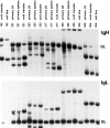
Evidence for the development of monoclonal/biclonal B lineage tumors in aging BALB-gld mice. Top and bottom panels show Southern blot analyses of genomic DNA isolated from the LN of representative 8 to 12 mo old BALB-gld mice bearing primary tumors, and from the LN of C.B-17-scid mice bearing the corresponding transplanted tumor (primary transplant). Data also are shown for splenic DNA from the primary 540 tumor and for DNA from the 311 cell line. In the top panel, DNA was digested with EcoRI and examined for rearrangement of the IgH locus with the pJ11 probe. In the bottom panel, DNA was digested with HindIII and examined for rearrangement of the IgL locus with the IVS probe. GL, indicates the position of the unrearranged germ-line IgH and IgL bands in BALB-gld kidney DNA (lane 19).
Figure 2
Figure 2
Evidence for the development of monoclonal/biclonal B lineage tumors in aging C3H-gld mice. Panels show Southern blot analyses of genomic DNA for IgH (top) and IgL (bottom) gene rearrangements (see Fig. 1 legend for details). DNA samples isolated from the spleen and LN of C3H-gld bearing primary tumors, from the LN of C3H-scid mice bearing primary tumor transplants and from cell lines are compared. GL indicates the position of the germline IgH and IgL bands.
Figure 3
Figure 3
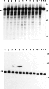
Clonal B cell outgrowths are not detected in the spleens of 12– 15-mo-old Balb/c+/+ mice. Panels show Southern blot analyses of genomic DNA for IgH (top) and IgL (bottom) gene rearrangements. DNA was isolated from the spleens of 12–15-mo-old Balb/c+/+ (lanes 1–11) or from Balb/c+/+ kidney (lane 12). GL indicates the position of the germline IgH and IgL bands. Data are representative of 25 mice analyzed.
Figure 4
Figure 4
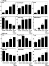
Age-related changes in the distribution of T and B cell subsets in the LN of C3H-gld and BALB-gld mice. The percentage of each lymphocyte subset (identified in top right hand corner) was determined by 3-color FACS® analysis of LN cells from individual BALB-gld and C3H-gld mice aged 4–5 mo, 7–10 mo, and >11 mo. Data is shown for all mice >11 mo and mice >11 mo with tumors. The proportions of B220+ DN T cells were determined by staining cells with PE-labeled anti-CD4 and anti-CD8, Tri-color-conjugated anti-CD45(B220), and FITC-labeled anti-TCR-α/β. The histograms represent the mean plus SE of values for 11 to 68 C3H-gld mice (top six panels) and 11–23 BALB-gld mice (bottom six panels).
Figure 5
Figure 5
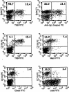
Age-related accumulation of B220+CD19+ThB+kappa+ CD23 B cells in gld mice. LN cells from a 12-mo-old BALB-gld mouse with no detectable tumor were stained with the reagents indicated. Values inside the quadrants represent the proportions of positive cells. Two thirds of the CD19+ B cells were Mac-1 and one third expressed low levels of Mac-1.
Figure 6
Figure 6
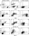
Phenotype of the C3H-gld tumor, 217 in vivo. Panels on the right hand side show FACS® data for LN cells from the mouse bearing the primary tumor 217. The adjacent panels show corresponding data for LN cells from C3H-scid mice bearing the primary (center) and secondary (right) 217 transplants. Cells were stained with the reagents indicated and analyzed on the FACScan®. The values inside each quadrant represent the percentage of reactive cells.
Figure 7
Figure 7
Cell surface marker expression on in vitro-propagated cell lines established from the C3H-gld tumor, 217, and the BALB-gld tumor, 311. Cells were stained with the antibodies indicated (heavy lines) or with a non-reactive FITC- or PE-conjugated control Ab (light lines) as described in Materials and Methods.
Figure 8
Figure 8
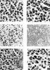
Malignant and nonmalignant lymphoid infiltrates in the tissues of aging gld mice. A illustrates the selective accumulation of DN T cells with characteristically stippled chromatin in a section of LN from a 4-mo-old C3H-gld mouse (H&E, original magnification 600×). B shows a typical example of the loss of the DN T cell population and the accumulation of nonmalignant plasma cells in the LN of a tumor-free 12-mo-old C3H-gld mouse (H&E, original magnification 600×). C and D show the morphologic heterogeneity of the cellular infiltrates in the spleen (C, original magnification 200×) and kidney (D, original magnification 100×) of the C3H-gld mouse bearing the primary 217 tumor. Panel E illustrates the extensive infiltration of the uterus with malignant plasmacytoid cells and plasma cells and non-malignant T lymphocytes in a C3H- scid mouse inoculated with the primary 217 tumor. F shows a LN section from a C3H-scid mouse bearing the second passage of tumor 217 in which the majority of the cells are monoclonal tumor cells (H&E, original magnification 600×). The mixture of immunoblasts, plasmablasts and plasma cells is typical of the morphologic heterogeneity associated with the malignant plasmacytoid gld lymphomas.
Figure 9
Figure 9
Evidence for somatically-acquired clonal MuLV proviral integrations in C3H-gld tumors. Southern blot of genomic DNA from primary tumors, scid-passaged tumors and tumor cell lines digested with PvuII and probed with a 32P-labeled ecotropic MuLV probe. DNA from C3H-gld kidney shows the position of the single hybridizing germline (GL) band present in normal C3H cells.
Figure 10
Figure 10
Somatically-acquired clonal MuLV proviral integrations in BALB-gld tumors. Southern blot analysis of PvuII-digested genomic DNA from BALB-gld primary tumors, scid transplants and the 311 cell line. The blot was hybridized with a 32P-labeled ecotropic MuLV probe. Control DNA samples from BALB-gld and C.B-17-scid kidneys show the position of the germline (GL) band.

Similar articles

Cited by

References

    1. Watanabe-Fukunada R, Brannan CI, Copeland NG, Jenkins NA, Nagata S. Lymphoproliferation disorder in mice explained by defects in Fas antigen that mediates apoptosis. Nature. 1992;356:314–317. - PubMed
    1. Takahashi T, Tanaka M, Brannan CI, Jenkins NA, Copeland NG, Suda T, Nagata S. Generalized lymphoproliferative disease in mice, caused by a point mutation in the Fas ligand. Cell. 1994;76:969–976. - PubMed
    1. Adachi M, Watanabe-Fukunaga R, Nagata S. Aberrant transcription caused by insertion of an early transposable element in an intron of the Fas antigen gene of lprmice. Proc Natl Acad Sci USA. 1993;90:1756–1760. - PMC - PubMed
    1. Suda T, Takahashi T, Goldstein P, Nagata S. Molecular cloning and expression of the Fas ligand, a novel member of the tumor necrosis factor family. Cell. 1993;75:1169–1178. - PubMed
    1. Yonehara S, Ishii A, Yonehara M. A cell-killing monoclonal antibody (anti-Fas) to a cell surface antigen co-downregulated with the receptor of tumor necrosis factor. J Exp Med. 1989;169:1747–1756. - PMC - PubMed