. 1998 Jun 1;187(11):1885-92.
doi: 10.1084/jem.187.11.1885.

Thalidomide costimulates primary human T lymphocytes, preferentially inducing proliferation, cytokine production, and cytotoxic responses in the CD8+ subset

Affiliations

Thalidomide costimulates primary human T lymphocytes, preferentially inducing proliferation, cytokine production, and cytotoxic responses in the CD8+ subset

P A Haslett et al. J Exp Med. .

Abstract

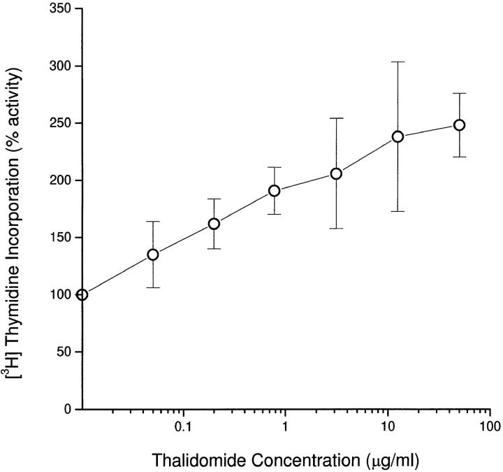
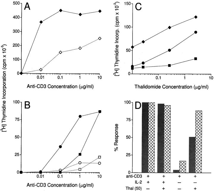
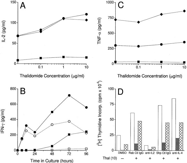
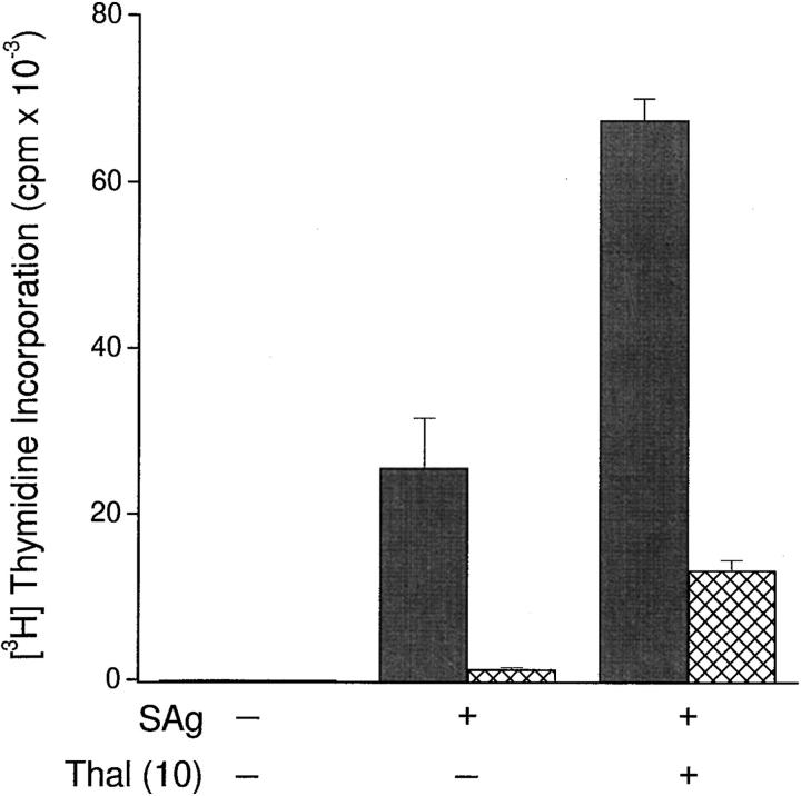
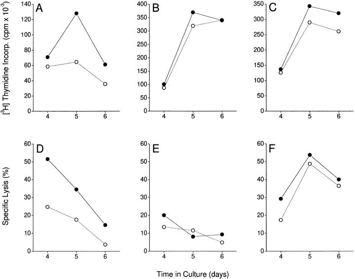
The efficacy of thalidomide (alpha-phthalimido-glutarimide) therapy in leprosy patients with erythema nodosum leprosum is thought to be due to inhibition of tumor necrosis factor alpha. In other diseases reported to respond to thalidomide, the mechanism of action of the drug is unclear. We show that thalidomide is a potent costimulator of primary human T cells in vitro, synergizing with stimulation via the T cell receptor complex to increase interleukin 2-mediated T cell proliferation and interferon gamma production. The costimulatory effect is greater on the CD8+ than the CD4+ T cell subset. The drug also increases the primary CD8+ cytotoxic T cell response induced by allogeneic dendritic cells in the absence of CD4+ T cells. Therefore, human T cell costimulation can be achieved pharmacologically with thalidomide, and preferentially in the CD8+ T cell subset.


Figures

Figure 1
Figure 1
Effect of thalidomide on mean proliferative responses of PBMCs from three healthy donors stimulated with 10 μg/ml immobilized anti-CD3. PBMCs (105/well) were cultured in triplicate at the indicated concentrations of thalidomide. [3H]Thymidine incorporation was measured for the last 12 h of 120-h cultures. Results are expressed as mean±SD. 100% activity on the y axis represents [3H]thymidine incorporation in the absence of thalidomide (DMSO-treated control).
Figure 2
Figure 2
(A) Effect of thalidomide (10 μg/ml) on the proliferative response of purified (>97% CD3+) T cells in the presence of increasing concentrations of immobilized anti-CD3. Filled symbols, Thalidomide-treated cultures. Open symbols, DMSO-treated controls. (B) Effect of thalidomide (10 μg/ml) on the proliferative response of highly purified positively selected CD4+ (squares) and CD8+ (circles) T cell subsets in the presence of increasing concentrations of immobilized anti-CD3. Filled symbols, Thalidomide-treated cells. Open symbols, DMSO-treated controls. (C) Effect of thalidomide on proliferation of purified bulk T cells (diamonds), CD4+ (squares), and CD8+ (circles) subsets in the presence of anti-CD3 at 0.1 μg/ml for bulk and CD8+, 10 μg/ml for CD4+ T cells. Symbols on the y axis represent responses in the absence of thalidomide (DMSO-treated control). T cells (105/well) were cultured in triplicate at the indicated concentrations of anti-CD3. [3H]Thymidine incorporation was measured for the last 12 h of 120-h cultures. (D) Lack of effect of thalidomide (Thal; 50 μg/ml) on CD4+ and CD8+ T cell proliferative responses to immobilized anti-CD3 (10 μg/ml) in the presence of exogenous r-hu IL-2 (10 U/ml). In the absence of exogenous IL-2, the enhancement by thalidomide of proliferation of CD8+ T cells is equivalent to that observed when exogenous IL-2 is present. Data expressed as percent activity of anti-CD3 plus IL-2–induced [3H]thymidine incorporation. T cells (105/well) were cultured in triplicate, and [3H]thymidine incorporation was measured for the last 12 h of 120-h cultures.
Figure 3
Figure 3
(A) Effect of thalidomide on IL-2 production by T cells stimulated with immobilized anti-CD3 (12 h). Diamonds, Bulk T cells. Squares, CD4+ T cells. Circles, CD8+ T cells. (B) Effect of thalidomide (10 μg/ml) on the kinetics of IFN-γ production by CD4+ (squares) and CD8+ (circles) T cells stimulated with immobilized anti-CD3. Filled symbols, Thalidomide-treated cells. Open symbols, DMSO-treated controls. (C) Lack of effect of thalidomide on TNF-α production by T cells stimulated with immobilized anti-CD3 (12 h). Symbols as in A. Cytokine concentrations were determined by ELISA of culture supernatants. No IL-4 was detected. (D) Effect of polyclonal rabbit anti– human IL-2 (anti-IL-2; 40 μg/ml) and polyclonal sheep anti–human IL-4 (anti-IL-4; 10 μg/ml) on thalidomide (Thal)-induced proliferation of bulk (white bars), CD4+ (gray bars), and CD8+ (hatched bars) T cells. Results for appropriate control antibodies (Rab Ctl IgG and Shp Ctl IgG) at the same concentrations are also shown. [3H]Thymidine incorporation was measured for the last 12 h of 120-h triplicate cultures.
Figure 4
Figure 4
Effect of thalidomide (Thal; 50 μg/ml) on CD4+ (gray bars) and CD8+ (hatched bars) T cell proliferative responses to SEA and SEB (combined, SAg: 10 ng/ml each) presented by autologous GAF ER− cells. Cultures were set up in triplicate in 96-well U-bottomed plates, with 105 T cells/well. ER−/T cell ratio was 1:1. [3H]Thymidine incorporation was measured for the last 12 h of 120-h cultures.
Figure 5
Figure 5
Effect of thalidomide on proliferative (top) and cytotoxic (bottom) T cell responses to allogeneic stimulation by blood-derived DCs. DC/T cell cocultures were set up at a ratio of 1: 30 for the durations indicated. Triplicate cultures of 105 purified CD8+ (A and D), CD4+ (B and E), and recombined CD4+/CD8+ (ratio 2:1; C and F) T cells were set up on day 0. For proliferation assays, 1 μCi [3H]thymidine was added for the last 12 h of culture. For cytotoxicity assays, 5 × 10³ 51Cr-labeled allogeneic monocytes (from the same donor as the DCs) were added to the cultures on the day of assay, and 51Cr release over 6 h was measured. Spontaneous release of 51Cr was <25% of the total. There was no detectable cytotoxicity against autologous (control) targets. Filled symbols, Thalidomide-treated cultures. Open symbols, DMSO-treated controls.

References

    1. Sheskin J. Thalidomide in the treatment of lepra reactions. Clin Pharmacol Ther. 1965;6:303–306. - PubMed
    1. Sampaio EP, Kaplan G, Miranda A, Nery JA, Miguel CP, Viana SM, Sarno EN. The influence of thalidomide on the clinical and immunologic manifestation of erythema nodosum leprosum. J Infect Dis. 1993;168:408–414. - PubMed
    1. Sampaio EP, Sarno EN, Galilly R, Cohn ZA, Kaplan G. Thalidomide selectively inhibits tumor necrosis factor α production by stimulated human monocytes. J Exp Med. 1991;173:699–703. - PMC - PubMed
    1. Waters MFR. An internally controlled double-blind trial of thalidomide in severe erythema nodosum leprosum. Lepr Rev. 1971;42:26–42. - PubMed
    1. Jacobson JM, Greenspan JS, Spritzler J, Ketter N, Fahey JL, Jackson JB, Fox L, Chernoff M, Wu AW, MacPhail LA, et al. Thalidomide for the treatment of oral aphthous ulcers in patients with human immunodeficiency virus infection. N Engl J Med. 1991;336:1487–1493. - PubMed

Publication types

MeSH terms