. 1998 Jun;111(6):857-73.
doi: 10.1085/jgp.111.6.857.

Cloning, expression, and characterization of the squid Na+-Ca2+ exchanger (NCX-SQ1)

Affiliations

Cloning, expression, and characterization of the squid Na+-Ca2+ exchanger (NCX-SQ1)

Z He et al. J Gen Physiol. 1998 Jun.

Abstract

We have cloned the squid neuronal Na+-Ca2+ exchanger, NCX-SQ1, expressed it in Xenopus oocytes, and characterized its regulatory and ion transport properties in giant excised membrane patches. The squid exchanger shows 58% identity with the canine Na+-Ca2+ exchanger (NCX1.1). Regions determined to be of functional importance in NCX1 are well conserved. Unique among exchanger sequences to date, NCX-SQ1 has a potential protein kinase C phosphorylation site (threonine 184) between transmembrane segments 3 and 4 and a tyrosine kinase site in the Ca2+ binding region (tyrosine 462). There is a deletion of 47 amino acids in the large intracellular loop of NCX-SQ1 in comparison with NCX1. Similar to NCX1, expression of NCX-SQ1 in Xenopus oocytes induced cytoplasmic Na+-dependent 45Ca2+ uptake; the uptake was inhibited by injection of Ca2+ chelators. In giant excised membrane patches, the NCX-SQ1 outward exchange current showed Na+-dependent inactivation, secondary activation by cytoplasmic Ca2+, and activation by chymotrypsin. The NCX-SQ1 exchange current was strongly stimulated by both ATP and the ATP-thioester, ATP gamma S, in the presence of F- (0.2 mM) and vanadate (50 microM), and both effects reversed on application of a phosphatidylinositol-4',5'-bisphosphate antibody. NCX1 current was stimulated by ATP, but not by ATP gamma S. Like NCX1 current, NCX-SQ1 current was strongly stimulated by phosphatidylinositol-4',5'-bisphosphate liposomes. In contrast to results in squid axon, NCX-SQ1 was not stimulated by phosphoarginine (5-10 mM). After chymotrypsin treatment, both the outward and inward NCX-SQ1 exchange currents were more strongly voltage dependent than NCX1 currents. Ion concentration jump experiments were performed to estimate the relative electrogenicity of Na+ and Ca2+ transport reactions. Outward current transients associated with Na+ extrusion were much smaller for NCX-SQ1 than NCX1, and inward current transients associated with Ca2+ extrusion were much larger. For NCX-SQ1, charge movements of Ca2+ transport could be defined in voltage jump experiments with a low cytoplasmic Ca2+ (2 microM) in the presence of high extracellular Ca2+ (4 mM). The rates of charge movements showed "U"-shaped dependence on voltage, and the slopes of both charge-voltage and rate-voltage relations (1,600 s-1 at 0 mV) indicated an apparent valency of -0.6 charges for the underlying reaction. Evidently, more negative charge moves into the membrane field in NCX-SQ1 than in NCX1 when ions are occluded into binding sites.


Figures

Figure 1
Figure 1
Stimulation of Ca2+-activated Cl current by polyvalent anions in an oocyte membrane patch. The pipette contains 80 mM and the cytoplasmic solution contains 10 mM Cl with NMG as the predominant cation. The free Ca2+ is buffered with 10 mM EGTA and membrane potential is 0 mV. (A) Application of a solution with 1 μM free Ca2+ activates outward Cl current, and additional application of 4 mM potassium phosphate (with 3 mM Mg2+, pH 7.0) enhances current by approximately fourfold over 1 min. The stimulatory effect reverses in just a few seconds on removal of phosphate. (B) Normalized stimulatory effects of other anions in the same protocol. “Basal” current corresponds to the current activated with 1 mM free Ca2+. From left to right, results are shown for 2 mM Mg-ATP, 2 mM Mg-AMPPNP, 2 mM ATP in the absence of Mg2+ from all solutions, 2 mM Mg-GTP, 1 mM Mg-GTPγS, 0.5 mM pyrophosphate (PP) in the absence of Mg2+ from all solutions, 4 mM p-nitrophenylphosphate (PnPP), 4 mM Mg-phosphoarginine (P-Arg), 4 mM F in the absence of Mg2+ from all solutions, and 2 mM EDTA in the absence of Mg2+ from all solutions.
Figure 2
Figure 2
Voltage-activated Na+ current in giant oocyte membrane patches. The pipette and the cytoplasmic solutions contain 40 mM Na+. (A) Depolarization from −40 to +40 mV activates an outward current over the course of 10 s, and the current deactivates without generating a significant tail current with signal filtering at 20 Hz. (B) Steady state current–voltage relation illustrating the steep voltage dependence of current activation. (C) Current–voltage relations under the same conditions, using 50-ms voltage steps as described in the text. After application of ATP (+ATP), the current is roughly doubled, runs down to less than control (after removal of ATP), and can be inhibited further with 20 μM Al3+ in the presence of 10 mM EGTA. The cytoplasmic solutions contained 0.1 mM F and 50 μM vanadate.
Figure 3
Figure 3
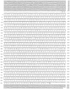
Nucleotide and deduced amino acid sequences of the squid Na+–Ca2+ exchanger clone, NCX-SQ1. Sequences have been submitted to GenBank under accession number U93214.
Figure 4
Figure 4
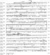
Amino acid comparison of the squid NCX-SQ1 and the canine NCX1 exchanger. Putative transmembrane segments, predicted by hydropathy analysis, are underlined and numbered. Highlighted in bold lettering are a potential signal peptidase site (SigPase), potential N-linked glycosylation sites (NXS/T), and potential phosphorylation sites (RTIK, protein kinase C; TRKLT, cAMP-dependent kinase and Ca2+/calmodulin-dependent kinase; DEHFY and DDEEEY, tyrosine kinase). The two potential phosphorylation sites marked with an asterisk are unique to NCX-SQ1. The endogenous exchanger inhibitory peptide (XIP) region and Exon A are shaded, and the binding domain for regulatory Ca2+ is boxed. The triple aspartate motifs involved in Ca2+ binding are in bold. Dots in the NCX1 sequence indicate amino acids identical to those of NCX-SQ1.
Figure 5
Figure 5
(left) Northern blot analysis of NCX-SQ1 RNA. mRNA (1 μg) from squid optical lobe (lane 1) and stellate ganglia (lane 2) was probed with a fragment of the NCX-SQ1 cDNA. (right) Western blot analysis of NCX-SQ1 protein. Protein from squid optical lobe vesicles (lane 1) and oocytes injected with water (lane 2) or cRNA for NCX-SQ1 (lane 3) was probed with an antibody raised against a histidine-tagged fusion protein fragment of NCX-SQ1.
Figure 5
Figure 5
(left) Northern blot analysis of NCX-SQ1 RNA. mRNA (1 μg) from squid optical lobe (lane 1) and stellate ganglia (lane 2) was probed with a fragment of the NCX-SQ1 cDNA. (right) Western blot analysis of NCX-SQ1 protein. Protein from squid optical lobe vesicles (lane 1) and oocytes injected with water (lane 2) or cRNA for NCX-SQ1 (lane 3) was probed with an antibody raised against a histidine-tagged fusion protein fragment of NCX-SQ1.
Figure 6
Figure 6
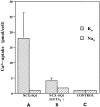
Functional expression of NCX-SQ1 in Xenopus oocytes. Oocytes injected with cRNA for NCX-SQ1 (A) or control (C), water-injected oocytes were assayed for Na+–Ca2+ exchanger activity. 45Ca2+ uptake into Na+ (90 mM)-loaded oocytes was measured in cells diluted into 45Ca2+-containing medium in the presence (extracellular K+) or absence of an outwardly directed Na+ gradient (extracellular Na+). In the middle pair of columns (B), 46 nl of a 100 mM EGTA solution was injected into the oocytes before loading the cells with Na+ to deplete intracellular Ca2+.
Figure 7
Figure 7
Outward Na+–Ca2+ exchange current of NCX-SQ1 in an excised oocyte membrane patch. Cytoplasmic solution with Na+ (40 mM) was applied and removed as indicated, first with 1.0 μM free cytoplasmic Ca2+, then with no cytoplasmic Ca2+, and finally with 5 μM free cytoplasmic Ca2+. The outward current, activated by application of Na+, inactivates partially over 10–50 s. The current is reduced by ∼50% in Ca2+-free solution. In the final sequence, α-chymotrypsin (1 mg/ml) was applied in the absence of cytoplasmic Ca2+. After complete activation by chymotrypsin, the current is insensitive to changes of cytoplasmic free Ca2+ in the micromolar range, and inactivation is abolished.
Figure 8
Figure 8
Stimulation of NCX1 outward exchange current in an oocyte patch by Mg-ATP. Cytoplasmic Na+-containing solution (40 mM) was applied and removed as indicated in the presence of 0.5 μM free cytoplasmic Ca2+. The solutions contain 0.2 mM F and 50 μM vanadate. (A) Application of 2 mM Mg-AMP-PNP is without effect, whereas 2 mM Mg-ATP stimulates the current to a magnitude somewhat greater than the initial peak current on application of cytoplasmic Na+. The stimulatory effect reverses partially over 2 min. (B) Application of 2 mM Mg-ATPγS has only a small stimulatory effect compared with 2 mM Mg-ATP, applied subsequently. The stimulatory effect reverses by ∼20% over 2 min, and it reverses almost completely in 1 min on application of PIP2 antibody (AB).
Figure 9
Figure 9
Stimulation of NCX-SQ1 outward exchange current in oocyte patches by Mg-ATP. Cytoplasmic solution with Na+ (40 mM) was applied and removed as indicated in the presence of 0.5 μM free cytoplasmic Ca2+. (A) Stabilization of stimulatory effect of ATP by F and vanadate. First, ATP was applied and removed in the absence of F and vanadate; the stimulatory effect of 2 mM Mg-ATP decays by ∼60% over 1 min after removal of ATP. Next, ATP was applied and removed in the presence of F and vanadate; after removal of ATP, the stimulatory effect is nearly stable for >1 min. (B) Reversal of the ATP effect by PIP2 antibody (PIP2-AB) in the presence of F and vanadate. Exchange current was activated by applying Na+, Mg-ATP was applied for 1 min, ATP was removed for 1 min, and finally PIP2-AB was applied. The stimulatory effect of ATP is stable after removal of ATP, but decays by ∼80% 90 s after application of antibody.
Figure 10
Figure 10
Stimulation of outward NCX-SQ1 Na+–Ca2+ exchange current by Mg-ATP-γ-S. (A) After activation of exchange current by applying Na+-containing solution, 2 mM Mg-AMP-PNP was applied for 1 min, resulting in no stimulatory effect. Then, 2 mM Mg-ATP-γ-S was applied, resulting in stimulation of the exchange current to above the initial peak obtained on applying Na+. The effect reversed on removal of nucleotide over ∼3 min. (B) After activating exchange current by applying 40 mM Na+-containing solution, the current was stimulated by applying 2 mM Mg-ATP-γ-S. On removal of the nucleotide, current is stable for 30 s. The stimulatory effect reverses in ∼1 min on application of PIP2 antibody (AB).
Figure 11
Figure 11
Stimulation of outward NCX-SQ1 Na+–Ca2+ exchange current by PIP2. (A) Current was activated by cytoplasmic Na+ and was then allowed to run down to <5 pA. PIP2 was applied and the outward current increased over 5 min to a magnitude more than twofold greater than the peak current on application of Na+. Current–voltage relations were taken just after application of PIP2 (1), after the maximum stimulatory effect was obtained (2), and after exchange current was turned off by removing cytoplasmic Na+ (3). (B) Current–voltage relation of the exchange current. The exchange current is defined by subtracting records before PIP2 application from those with PIP2 (2-1, •) and those after removing Na+ from those with Na+ plus PIP2 (2-3, ○). (C) Comparison of current transients obtained on activating exchange current before (control) and after (+PIP2) applying PIP2. The time constant (t) of inactivation increases from 3.9 to 8.2 s. Results are from a different patch.
Figure 12
Figure 12
Lack of effect of phosphoarginine (p-ARG) on NCX-SQ1 exchange current in an excised oocyte patch. After activation of the exchange current by applying Na+-containing solution, 5 mM phosphoarginine was applied with 3 mM Mg2+ (pH 7.0). There is no stimulatory effect, whereas application of 50 μM PIP2 strongly stimulates the exchange current to a magnitude more than twofold greater than the peak obtained on applying Na+ initially.
Figure 13
Figure 13
Outward NCX-SQ1 Na+–Ca2+ exchange current in a chymotrypsin-treated patch. The pipette solution contains 4 mM Ca2+ and no Na+; the cytoplasmic solution contains 10 mM EGTA and no Ca2+. (A) Current–voltage relations at the given Na+ concentrations from 5 to 90 mM. Data points for descending and ascending voltage steps show no hysteresis. (B) Cytoplasmic Na+ dependence of outward exchange current at +60 and −60 mV. The data points are fit to a Hill equation; the slope is 1.2 at +60 and 1.7 at −60 mV; the K50 is 27 mM at +60 mV and 24 mM at −60 mV.
Figure 14
Figure 14
Comparison of inward NCX1 and NCX-SQ1 Na+– Ca2+ exchange currents in chymotrypsin-treated patches. The pipette solution contains (mM): 120 Na+, 10 EGTA, 20 Cs+, 20 HEPES, 4 Mg2+, and no Ca2+ (pH 7.0 with NMG); the cytoplasmic solution contains 10 mM EGTA and no Na+. The inward current– voltage relations are defined by subtracting records with Ca2+ from records without Ca2+. In descending order, the current–voltage relations are with 0.2, 2, 10, and 300 μM Ca2+. (A) Current–voltage relations for NCX1. (B) Current–voltage relations for NCX-SQ1. Same batch of oocytes as in A. (C) Cytoplasmic Ca2+ dependence of the inward NCX1 exchange current at −150 and −30 mV. The K50, indicated by an arrow, is 2.5 μM at −30 mV and 4.2 μM at −150 mV. (D) Cytoplasmic Ca2+ dependence of the inward NCX-SQ1 exchange current at −150 and −30 mV. The K50 is 3.5 μM at −30 mV and 7.2 μM at −150 mV.
Figure 15
Figure 15
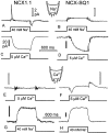
Identification of electrogenic reactions of NCX1 and NCX-SQ1 using concentration jumps. (A) Current transients recorded from NCX1-expressing patch when 40 mM cytoplasmic Na+ is applied and removed in the presence of 20 mM extracellular Na+. (B) Typical lack of current transients recorded from NCX-SQ1-expressing patch when 40 mM cytoplasmic Na+ is applied and removed in the presence of 20 mM extracellular Na+. (C) Inward NCX1 current activated when a solution with 5 μM free Ca2+ is applied as in A. (D) Inward NCX-SQ1 current activated when a solution with 5 μM free Ca2+ is applied as in B. (E) Typical lack of current transients for a Ca2+ jump to 5 μM free Ca2+ in NCX1- expressing patch; 50 μM extracellular Ca2+. (F) Current transients recorded from NCX-SQ1-expressing patch when a solution with 5 μM free Ca2+ is applied and removed in the presence of 50 μM extracellular Ca2+. (G) Outward current activated by applying 40 mM Na+ to an NCX1 patch with 50 μM extracellular Ca2+. (H) Outward current activated by applying 40 mM Na+ to an NCX1 patch with 50 μM extracellular Ca2+. See text for details.
Figure 16
Figure 16
Charge movements related to Ca2+ transport by NCX-SQ1 Na+–Ca2+ exchanger. (A) In the presence of 4 mM extracellular Ca2+, charge records were taken with and without 2 μM cytoplasmic free Ca2+. The holding potential was −40 mV, and potential was stepped for 1 ms to different values in 40-mV steps. Membrane current is the first derivative of these records. Signals were essentially flat at the same amplification in control oocyte patches. (B) Voltage dependence of the Ca2+-dependent charge movements recorded in A. The Boltzmann slope (q) of the fitted Boltzmann function was 0.46. (C) Voltage dependence of rates of the charge movements, determined by fitting the charge transients to single exponential functions. The Boltzmann slope (q) of the fitted sum of two exponentials is 0.59. See text for details.

Similar articles

Cited by

References

    1. Baker PF, McNaughton PA. Kinetics and energetics of calcium efflux from intact squid axons. J Physiol (Camb) 1976;259:104–114. - PMC - PubMed
    1. Baud C, Kado RT. Induction and disappearance of excitability in the oocyte of Xenopus laevis: a voltage-clamp study. J Physiol (Camb) 1984;356:275–289. - PMC - PubMed
    1. Chomczynski P, Sacchi N. Single-step method of RNA isolation by acid guanidinium thiocyanate-phenol-chloroform extraction. Anal Biochem. 1987;162:156–159. - PubMed
    1. Collins A, Somlyo A, Hilgemann DW. The giant cardiac membrane patch method: stimulation of outward Na/Ca exchange current by MgATP. J Physiol (Camb) 1992;454:37–57. - PMC - PubMed
    1. DiPolo R. The effect of ATP on Ca2+efflux in dialyzed squid giant axons. J Gen Physiol. 1974;64:503–517. - PMC - PubMed

Publication types

Substances