

Antimicrobial management of presumed microbial keratitis: guidelines for treatment of central and peripheral ulcers
- PMID: 9613378
- PMCID: PMC1722498
- DOI: 10.1136/bjo.82.2.137
Antimicrobial management of presumed microbial keratitis: guidelines for treatment of central and peripheral ulcers
Abstract
Aims: To determine the quantitative relation between the major risk factors for microbial keratitis of previous ocular surface disease and contact lens wear and central and peripheral infiltration, often associated with ulceration, in order to establish a rational chemotherapeutic management algorithm.
Methods: Data from 55 patients were collected over a 10 month period. All cases of presumed microbial keratitis where corneal scrapes had been subjected to microbiological examination were included. Risk factor data and laboratory outcome were recorded. Antimicrobial regimens used to treat each patient were documented.
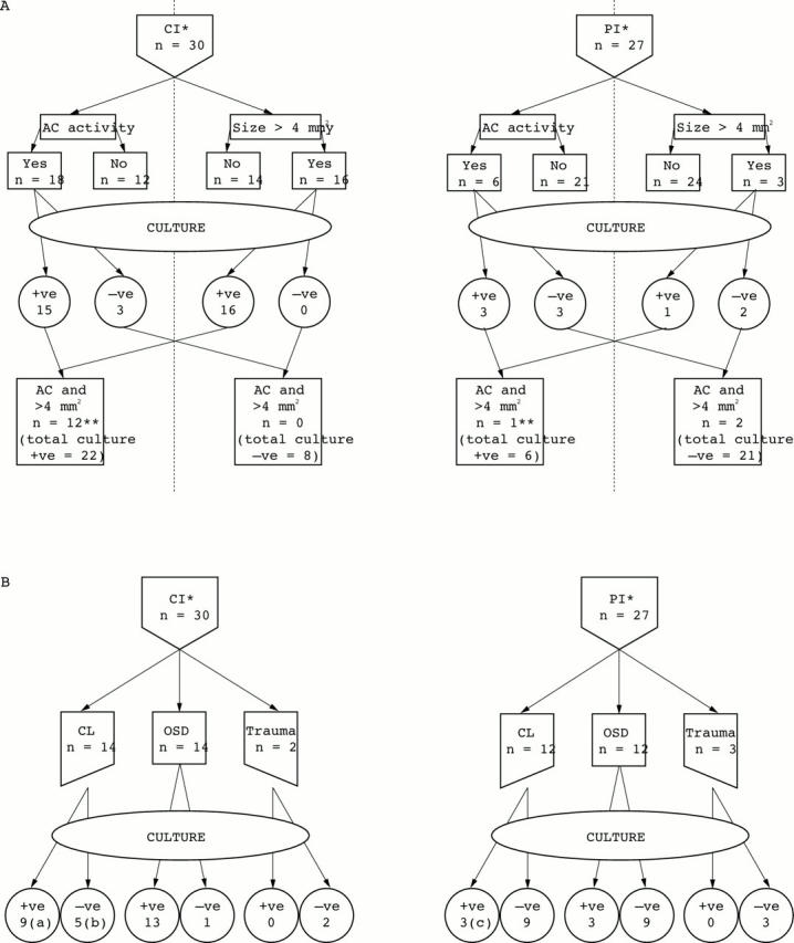
Results: 57 episodes of presumed microbial keratitis were identified from 55 patients, 24 male and 31 female. There were 30 central infiltrates and 27 peripheral infiltrates of which 28 were culture positive (73% of central infiltrates, 22% of peripheral infiltrates). 26 patients had worn contact lenses of whom 12 had culture positive scrapes (9/14 for central infiltrates, 3/12 for peripheral infiltrates). 31 patients had an ocular surface disease of whom five previous herpes simplex virus keratitis patients developed secondary bacterial infection. Anterior chamber activity and an infiltrate size > or = 4 mm2 were more common with culture positive central infiltrates than peripheral infiltrates (chi 2 test = 11.98, p < 0.001).
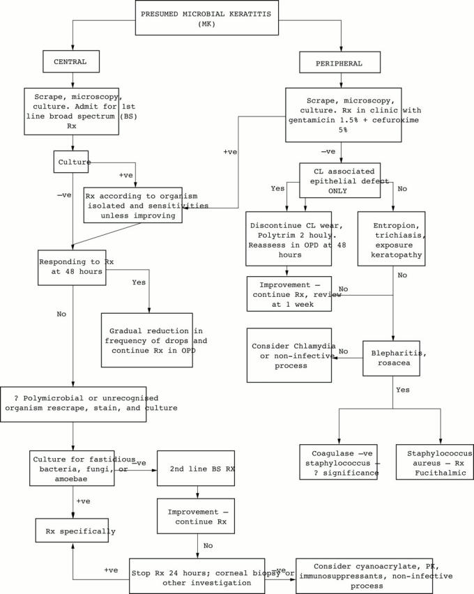
Conclusions: Predisposing factors for "presumed" microbial keratitis, either central or peripheral, were: ocular surface disease (26/57 = 45.6%), contact lens wear (26/57 = 45.6%), and previous trauma (5/57 = 8.8%). Larger ulceration (> or = 4 mm2) with inflammation was more often associated with positive culture results for central infiltration. None of these four variables (contact lens wear, ocular surface disease, ulcer size, anterior chamber activity) were of intrinsic value in predicting if a peripheral infiltrate would yield identifiable micro-organisms. Successful management of presumed microbial keratitis is aided by a logical approach to therapy, with the use of a defined algorithm of first and second line broad spectrum antimicrobials, for application at each stage of the investigative and treatment process considering central and peripheral infiltration separately.
Figures
References
Publication types
MeSH terms
LinkOut - more resources
Full Text Sources
Medical