. 1998 Jun;9(6):1425-35.
doi: 10.1091/mbc.9.6.1425.

Identification of osteopontin as a novel ligand for the integrin alpha8 beta1 and potential roles for this integrin-ligand interaction in kidney morphogenesis

Affiliations
Free PMC article

Identification of osteopontin as a novel ligand for the integrin alpha8 beta1 and potential roles for this integrin-ligand interaction in kidney morphogenesis

S Denda et al. Mol Biol Cell. 1998 Jun.
Free PMC article

Abstract

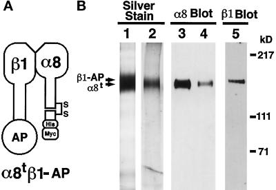
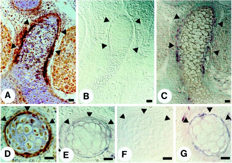
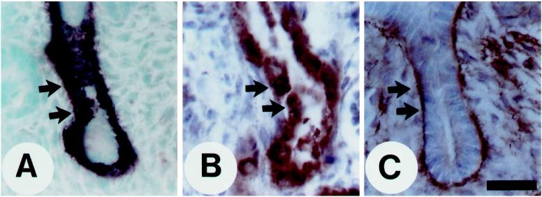
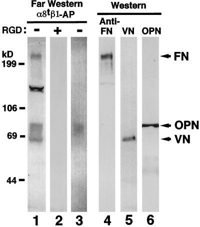
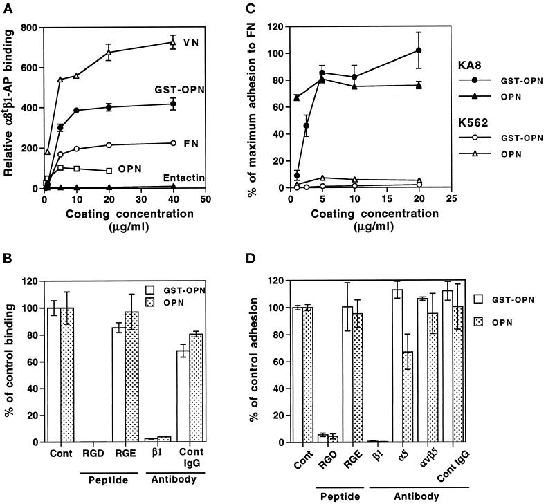
Epithelio-mesenchymal interactions during kidney organogenesis are disrupted in integrin alpha8 beta1-deficient mice. However, the known ligands for integrin alpha8 beta1-fibronectin, vitronectin, and tenascin-C-are not appropriately localized to mediate all alpha8 beta1 functions in the kidney. Using a method of general utility for determining the distribution of unknown integrin ligands in situ and biochemical characterization of these ligands, we identified osteopontin (OPN) as a ligand for alpha8 beta1. We have coexpressed the extracellular domains of the mouse alpha8 and beta1 integrin subunits as a soluble heterodimer with one subunit fused to alkaline phosphatase (AP) and have used the alpha8 beta1-AP chimera as a histochemical reagent on sections of mouse embryos. Ligand localization with alpha8 beta1-AP in developing bone and kidney was observed to be overlapping with the distribution of OPN. In "far Western" blots of mouse embryonic protein extracts, bands were detected with sizes corresponding to fibronectin, vitronectin, and unknown proteins, one of which was identical to the size of OPN. In a solid-phase binding assay we demonstrated that purified OPN binds specifically to alpha8 beta1-AP. Cell adhesion assays using K562 cells expressing alpha8 beta1 were used to confirm this result. Together with a recent report that anti-OPN antibodies disrupt kidney morphogenesis, our results suggest that interactions between OPN and integrin alpha8 beta1 may help regulate kidney development and other morphogenetic processes.


Figures

Figure 1
Figure 1
Purification of soluble truncated integrin α8β1 heterodimer and α8 monomer. (A) Schematic representation of the soluble α8tβ1-AP heterodimer. Mature integrin α8 and β1 subunits consist of extracellular, transmembrane, and cytoplasmic domains. The truncated α8 (α8t) consists of the entire extracellular domain of mouse α8 cDNA with c-terminal (His)6 and c-myc epitope tags. The β1-AP chimera (β1-AP) consists of the entire extracellular domain of β1 fused in frame to human placental AP. α8tβ1-AP is a heterodimer of α8t and β1-AP. (B) The purified α8tβ1-AP heterodimer (lanes 1, 3, and 5) and α8t monomer (lanes 2 and 4) were separated by electrophoresis in 6% polyacrylamide gels in nonreducing conditions. The gels were either silver stained (lanes 1 and 2) or transferred to nitrocellulose membranes and probed with anti-α8 antiserum (lanes 3 and 4) or anti-β1 mAb 9EG7 (lane 5). In lane 1, both α8t and β1-AP bands are within the overexposed band shown. The purified α8t monomer (lane 2) was used as an antigen to prepare the rabbit polyclonal anti-mouse α8 antibody.
Figure 2
Figure 2
Colocalization of α8tβ1-AP binding sites and OPN in the regions of bone morphogenesis. Paraffin sections of E16.5 (A–C) and E13.5 (D–G) mouse embryos were incubated with indicated reagents: (A) anti-OPN peptide antiserum; (B) control conditioned medium containing nonfused, secreted AP; (C and E) α8tβ1-AP; (D) anti-GST-mouse OPN IgG; (F) α8tβ1-AP in the presence of 50 μg/ml GRGDSP peptide; and (G) α8tβ1-AP in the presence of 50 μg/ml GRGESP peptide. Arrowheads indicate the bony collar. Bar, 10 μm.
Figure 3
Figure 3
Colocalization of α8tβ1-AP binding sites, OPN, and the integrin α8 subunit in the developing kidney. Paraffin sections (A and C) and frozen sections (B) of an E16.5 kidney were incubated with α8tβ1-AP (A), anti-OPN peptide antiserum (B), and affinity-purified anti-mouse integrin α8 subunit IgG (C). The panels show a cross-section through the ureter epithelium, and the arrows point to the border between the epithelium and surrounding mesenchyme. Note that the section is at a level below the tips of the ureter; thererfore, no condensing mesenchymal cells are visible. Bar, 50 μm.
Figure 4
Figure 4
RGD-dependent binding of α8tβ1-AP to proteins including OPN in mouse embryo extract. Fifty micrograms of protein per lane of the extract from E13.5 embryo (lanes 1, 2, and 4–6) and immunoprecipitates from E16.5 kidney extract with OPN antiserum pp69 (lane 3) were separated electrophoretically on a 7.5% polyacrylamide gel in reducing conditions and analyzed by far Western (lanes 1–3) and Western (lanes 4–6) blotting. For far Western blotting of the E13.5 extract, membrane strips were incubated with α8tβ1-AP in the absence (lane 1) or presence (lane 2) of 50 μg/ml GRGDSP peptide. For Western blotting, the membrane strips were probed with anti-FN IgG (lane 4), anti-VN IgG (lane 5), or anti-OPN mAb MPIIIB10 (lane 6). The integrin-AP chimera bound to protein bands of 240, 130, and 65–85 kDa in the extract (lane 1) and ∼80 kDa in the anti-OPN immunoprecipitates (lane 3). This 80-kDa band comigrates with the band detected by OPN Western blot (lane 6).
Figure 5
Figure 5
Binding of α8tβ1-AP and cells expressing α8β1 to known ligands and to purified native OPN and recombinant GST-OPN. (A and B) Solid-phase binding assays were performed with α8tβ1-AP in the presence of 1 mM MnCl2. (A) α8tβ1-AP binding to entactin, VN, FN, native OPN, and GST-OPN. Plates were coated with indicated concentrations of purified proteins. The absorbance values were normalized using the absorbance value for binding to substrata coated with 10 μg/ml FN120. Results demonstrate that α8tβ1-AP binds to OPN and GST-OPN as well as to FN and VN. (B) Effects of peptides and an inhibitory anti-β1 antibody on α8tβ1-AP binding to native OPN and GST-OPN. Wells coated with 5 μg/ml OPN or 10 μg/ml GST-OPN were incubated with α8tβ1-AP with additions indicated: (Cont) no addition; (RGD) 0.1 μg/ml GRGDSP peptide; (RGE) 0.1 μg/ml GRGESP peptide; (β1) 100 μg/ml anti-integrin β1 mAb HA2/11; and (Cont IgG) 100 μg/ml control IgG. The absorbance was quantified as the percentage of control binding (no addition). (C) Cell adhesion to native OPN and GST-OPN. Cell adhesion assays were carried out with KA8 and K562 cells on plates coated with indicated concentrations of OPN and GST-OPN in the presence of 1 mM MnCl2. The absorbance was quantified as the percentage of maximum adhesion to FN coated at 5 μg/ml. Note that only KA8 cells adhered to OPN and GST-OPN. (D) Effects of peptides and anti-integrin mAbs on KA8 cell adhesion to OPN and GST-OPN. KA8 cell adhesion assays were carried out on OPN and GST-OPN (coated at 5 μg/ml) in the presence of 1 mM MnCl2 with additions as indicated: (Cont) no addition; (RGD) 10 μg/ml GRGDSP peptide; (RGE) 10 μg/ml GRGESP peptide; (β1) anti-β1 mAb AIIB2; (α5) anti-α5 mAb B1E5; (αvβ5) anti-αvβ5 mAb P1F6; and (Cont IgG) 100 μg/ml control IgG. Adhesion with no addition (Cont) was set at 100%. Note that KA8 cell adhesion to OPN and GST-OPN was RGD sensitive and was blocked by the anti-β1 mAb. Data represent mean ± SD of triplicate wells.
Figure 6
Figure 6
Cell-spreading assay. In the presence of 1 mM MnCl2, K562 cells (A, C, E, and G) and KA8 cells (B, D, F, and H) were plated onto different substrata: (A and B) BSA; (C and D) FN (coated at 5 μg/ml); (E and F) purified native OPN (5 μg/ml); and (G and H) recombinant GST-OPN (5 μg/ml). Note that both cell types spread on FN, but only KA8 cells spread on native OPN and recombinant GST-OPN. Bar, 100 μm.

Similar articles

Cited by

References

    1. Aufderheide E, Chiquet-Ehrismann R, Ekblom P. Epithelial-mesenchymal interactions in the developing kidney lead to expression of tenascin in the mesenchyme. J Cell Biol. 1987;105:599–608. - PMC - PubMed
    1. Bossy B, Bossy-Wetzel E, Reichardt LF. Characterization of the integrin alpha 8 subunit: a new integrin beta 1-associated subunit, which is prominently expressed on axons and on cells in contact with basal laminae in chick embryos. EMBO J. 1991;10:2375–2385. - PMC - PubMed
    1. Chen J, Zhang Q, McCulloch CAG, Sodek J. Immunohistochemical localization of bone sialoprotein in foetal procine bone tissues: comparisons with secreted phosphoprotein 1 (SPP-1, osteopontin) and SPARC (osteonectin) Histochem J. 1991;23:281–289. - PubMed
    1. Cheng H-J, Flanagan JG. Identification and cloning of ELF-1, a developmentally expressed ligand for the Mek4 and Sek receptor tyrosine kinases. Cell. 1994;79:157–168. - PubMed
    1. Clover J, Dodds RA, Gowen M. Integrin subunit expression by human osteoblasts and osteoclasts in situ and in culture. J Cell Sci. 1992;103:267–271. - PubMed

Publication types

MeSH terms