. 1998 Jun;9(6):1479-94.
doi: 10.1091/mbc.9.6.1479.

Effects of age on the posttranscriptional regulation of CCAAT/enhancer binding protein alpha and CCAAT/enhancer binding protein beta isoform synthesis in control and LPS-treated livers

Affiliations
Free PMC article

Effects of age on the posttranscriptional regulation of CCAAT/enhancer binding protein alpha and CCAAT/enhancer binding protein beta isoform synthesis in control and LPS-treated livers

C C Hsieh et al. Mol Biol Cell. 1998 Jun.
Free PMC article

Abstract

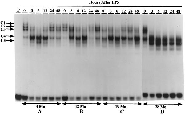
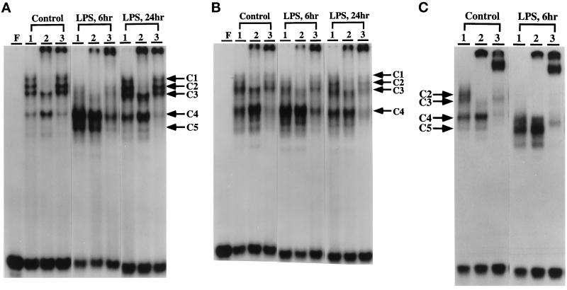
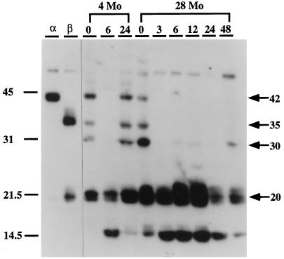
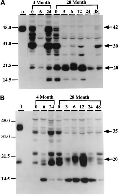
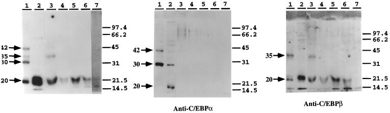
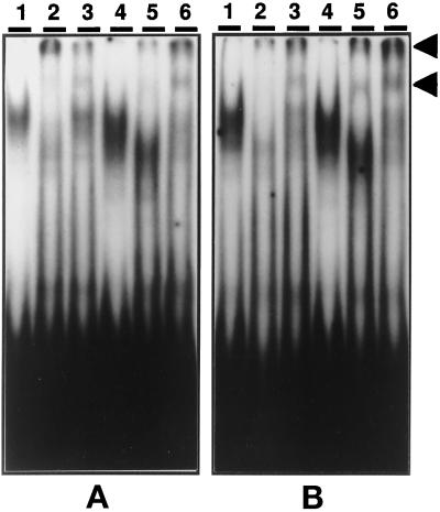
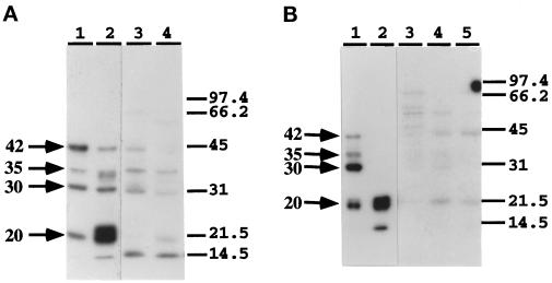
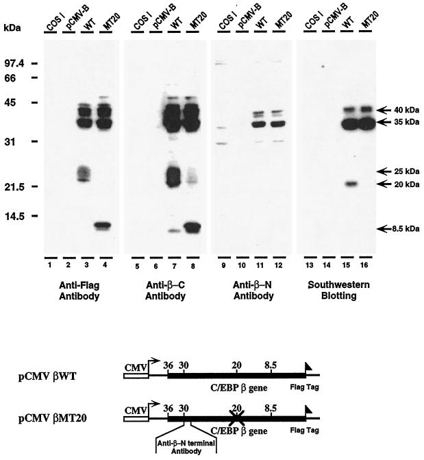
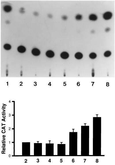
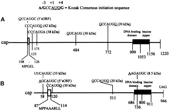
The CCAAT/enhancer binding protein alpha (C/EBPalpha) and CCAAT/enhancer binding protein beta (C/EBPbeta) mRNAs are templates for the differential translation of several isoforms. Immunoblotting detects C/EBPalphas with molecular masses of 42, 38, 30, and 20 kDa and C/EBPbetas of 35, 20, and approximately 8.5 kDa. The DNA-binding activities and pool levels of p42(C/EBPalpha) and p30(C/EBPalpha) in control nuclear extracts decrease significantly whereas the binding activity and protein levels of the 20-kDa isoforms increase dramatically with LPS treatment. Our studies suggest that the LPS response involves alternative translational initiation at specific in-frame AUGs, producing specific C/EBPalpha and C/EBPbeta isoform patterns. We propose that alternative translational initiation occurs by a leaky ribosomal scanning mechanism. We find that nuclear extracts from normal aged mouse livers have decreased p42(C/EBPalpha) levels and binding activity, whereas those of p20(C/EBPalpha) and p20(C/EBPbeta) are increased. However, translation of 42-kDa C/EBPalpha is not down-regulated on polysomes, suggesting that aging may affect its nuclear translocation. Furthermore, recovery of the C/EBPalpha- and C/EBPbeta-binding activities and pool levels from an LPS challenge is delayed significantly in aged mouse livers. Thus, aged livers have altered steady-state levels of C/EBPalpha and C/EBPbeta isoforms. This result suggests that normal aging liver exhibits characteristics of chronic stress and a severe inability to recover from an inflammatory challenge.


Figures

Figure 1
Figure 1
A time course of DNA binding to the C/EBP-binding site (APRE) of the mouse AGP-1 promoter with nuclear proteins from livers of young and aged mice treated with LPS. EMSAs were performed using 3 μg of nuclear proteins isolated from fresh livers of control (0) or LPS-injected C57BL/6 male mice aged 4 mo (A), 12 mo (B), 19 mo (C), and 28 mo (D). LPS (10 μg) was injected at time 0, and mice were killed and nuclear proteins prepared at the times indicated at the top of each lane. The double-stranded oligonucleotide corresponding to the APRE of AGP-1 promoter was labeled with [γ-32P]ATP and T4 polynucleotide kinase. The DNA–protein complexes are indicated as C1, C2, C3, C4, and C5. Lane F contains the free DNA probe.
Figure 2
Figure 2
Mobility supershift analyses of the DNA–protein complexes formed with the APRE of the AGP-1 promoter. Supershift assays were done as described in MATERIALS AND METHODS to demonstrate that the nuclear proteins involved in the formation of DNA–protein complexes with the AGP-1 APRE are members of the C/EBP family. Monospecific antibodies against synthetic peptides unique to the C/EBPα and C/EBPβ isoforms were used. Lanes F, free oligonucleotide; lanes 1, no antibody was added to the reaction mixture; lanes 2, mouse anti-C/EBPα; lanes 3, mouse anti-C/EBPβ. Nuclear extracts were prepared 0, 6, and 24 h after LPS injection as indicated. Mice 4, 19, and 28 mo of age were used. The positions of the DNA–protein complexes are noted as C1–C5; the bands formed by supershifting with C/EBPα or C/EBPβ antibody are indicated by the arrows. (A) 4 mo. (B) 19 mo. (C) 28 mo.
Figure 3
Figure 3
Southwestern blot analysis of liver nuclear proteins that bind to the APRE of the AGP-1 gene after LPS treatment. C57BL/6 mice (4- and 28-mo-old males) were injected with 10 μg of LPS and then killed 3, 6, 12, 24, or 48 h after injection. Nuclear proteins (30 μg) prepared from fresh livers were subjected to SDS-PAGE, blotted onto Westran PVDF membranes, and probed with 106 cpm/ml of 32P-labeled APRE oligonucleotide as described in MATERIALS AND METHODS. Locations of molecular size standards (kDa) are shown on the left. Lanes α and β contain nuclear extracts from COS-1 cells transfected with expression plasmids for C/EBPα and C/EBPβ, respectively, as described in MATERIALS AND METHODS.
Figure 4
Figure 4
Western blot analysis of the levels of C/EBPα and C/EBPβ isoforms in liver nuclei in response to LPS treatment. Nuclear extracts (30 μg) from control (0) and LPS-injected (3, 6, 12, 24, and 48 h postinjection) C57BL/6 mice (4- and 28-mo-old males) were loaded in each lane and subjected to ECL-Western immunoblot analyses as described in MATERIALS AND METHODS. Immunoblots were incubated with monospecific polyclonal antibodies against C/EBPα (A) or C/EBPβ (B) or preimmune serum as a control (unpublished data). (A) Anti-C/EBPα. (B) Anti-C/EBPβ. Lanes α and β represent nuclear extracts from COS-1 cells transfected with C/EBPα or C/EBPβ expression vectors and were used as standards for each protein. No bands were detected with preimmune serum with liver nuclear extracts or COS-1 nuclear proteins (unpublished data). Positions of molecular mass standards (kDa) are indicated on the left.
Figure 5
Figure 5
Identification of the C/EBP proteins that interact with APRE oligonucleotide to form C1, C2, C3, C4, and C5 complexes in livers of aged mice in response to LPS. The C1–C3, C4, and C5 complexes formed with control or LPS-treated (6 h) nuclear extracts from 28-mo-old C57BL/6 mice were resolved by preparative EMSA. The bands were excised from the gel, eluted, concentrated, and subjected to Southwestern blot and to ECL-Western immunoblot analyses as described in Ref. 3. (A) Southwestern analysis of liver nuclear proteins from 28-mo-old mice that form the C1–C3, C3, C4, and C5 DNA–protein complexes with the APRE oligonucleotide. Lane 1, control NE; lane 2, LPS-treated NE; lane 3, C1–C3 complexes formed with control NE; lane 4, C4 complex formed with control NE. Lanes 5 and 6, C3 and C4 complexes, respectively, formed with mouse liver NE after 6 h of LPS treatment. Lane 7, C5 complex 6 h after LPS treatment. (B and C) Western blot analysis using anti-C/EBPα and anti-C/EBPβ and the same nuclear extracts and complexes as in A.
Figure 6
Figure 6
EMSA and supershift analysis of mouse liver polysomal proteins that bind to the APRE oligonucleotide. Liver polysomes from young (A, 4-mo-old) and aged (B, 28-mo-old) control and LPS-treated mice were prepared as described in MATERIALS AND METHODS. The [32P]APRE oligonucleotide was prepared as described in MATERIALS AND METHODS. (A) Liver polysomal proteins from 4-mo-old mice; lanes 1–3, control; lane 2, anti-C/EBPα; lane 3, anti-C/EBPβ; lanes 4–6, liver polysomal proteins from 4-mo-old LPS-treated mice; lane 4, no antibody; lane 5, anti-C/EBPα; lane 6, anti-C/EBPβ. (B) Liver polysomal proteins from 28-mo-old mice; lanes 1–3, control; lane 2, anti-C/EBPα; lane 3, anti-C/EBPβ; lanes 4–6, LPS treated; lane 4, no antibody; lane 5, anti-C/EBPα; lane 6, anti-C/EBPβ; lane F, free probe.
Figure 7
Figure 7
Southwestern blot analysis of the mouse liver polysomal proteins from young (A, 4 mo) and aged (B, 28 mo) old livers. (A) Lane 1, control NE; lane 2, LPS-treated NE; lane 3, control polysomal proteins; lane 4, polysomal proteins 3 h after LPS treatment. (B) Lane 1, control NE; lane 2, LPS-treated NE; lane 3, control polysomal proteins; lane 4, polysomal proteins 3 h after LPS treatment; lane 5, polysomal proteins 6 h after LPS treatment.
Figure 8
Figure 8
The transcription and translation of wild-type and mutant pCMV-C/EBPβ expression vectors in COS-1 cells. Western blot analysis of the C/EBPβ isoforms synthesized in COS-1 cells transfected with wild-type pCMV-C/EBPβ and pCMV-C/EBPβ mutated at the 20-kDa AUG start site. The AUG was mutated to TTG. The expression vectors contain a Flag tag at their C-terminal ends. Western blot analyses were performed on COS-1 nuclear extracts using anti-Flag (lanes 1–4), anti-C-terminal C/EBPβ-antibodies (lanes 5–8); and anti-N-terminal C/EBPβ antibody (lanes 9–13). Southwestern blot analysis was performed on the COS-1 nuclear extracts used for the immunoblot analyses (lanes 13–16). Lanes 1, 5, 9, and 13, control COS-1 nuclear extract; lanes 2, 6, 10, and 14, nuclear extracts of COS-1 cells transfected with expression vector lacking C/EBPβ sequences (pCMV-B); lanes 3, 7, 11, and 15, nuclear extracts of COS-1 cells transfected with wild-type pCMV-C/EBPβ; lanes 4, 8, 12, and 16, nuclear extracts from COS-1 cells transfected with pCMV-C/EBPβ mutated at 20-kDa AUG start site. Maps of the C/EBPβ expression vectors used in these experiments are shown below the autoradiogram.
Figure 9
Figure 9
The transactivation of pAPRE-CAT expression by p20C/EBPβ. COS-1 cells were cotransfected with pMSV-C/EBPβ20-kDa and pAPRE-CAT expression vectors to determine whether expression of the 20-kDa C/EBPβ isoform could activate expression of pAPRE-CAT. A typical autoradiogram depicting chloramphenicol acetyl transferase activity in response to increasing expression of 20-kDa C/EBPβ is shown in the upper panel. Lane 1, the pSV2-CAT expression vector minus the APRE sequences; lane 2, pAPRE-CAT; lanes 3–5, pAPRE-CAT + 1, 2, and 3 μg of pMSV without the 20-kDa C/EBPβ sequences, respectively; lanes 6–8, pAPRE-CAT + 1, 2, and 3 μg of pMSV-C/EBPβ20-kDa, respectively. A presentation of the data from three separate experiments is shown in the lower panel.
Figure 10
Figure 10
A map of the C/EBPα (A) and C/EBPβ (B) mRNAs. The positions of the AUG initiation sites within each mRNA and the Kozak sequences relative to the A (+1) in each AUG are indicated. The numbers indicate the position of the As in each initiation codon. The molecular masses of isoforms that can be formed by initiation at the designated AUG sites are depicted in parentheses behind each Kozak sequence. The MPGEL is the amino acid sequence of the C/EBPα sORF and MPPAAARLL is the amino acid sequence of the C/EBPβ sORF.

References

    1. Alam T, An MR, Mifflin RC, Hsieh C-C, Ge X, Papaconstantinou J. Trans-activation of the α1-acid-glycoprotein gene acute phase responsive element by multiple isoforms of C/EBP and glucocorticoid receptor. J Biol Chem. 1993;268:15681–15688. - PubMed
    1. Alam T, Papaconstantinou J. Interaction of acute-phase-inducible and liver enriched nuclear factors with the promoter region of the mouse α1-acid glycoprotein gene-1. Biochemistry. 1992;31:1928–1936. - PubMed
    1. An MR, Hsieh C-C, Reisner PD, Rabek JR, Scott SG, Kuninger DT, Papaconstantinou J. Evidence for posttranscriptional regulation of C/EBPα and C/EBPβ isoform expression during the lipopolysaccharide-mediated acute-phase response. Mol Cell Biol. 1996;16:2295–2306. - PMC - PubMed
    1. Baumann H, Jahreis GP, Morella KK. Interaction of cytokine- and glucocorticoid-response elements of acute-phase plasma protein genes. J Biol Chem. 1990;265:22275–22281. - PubMed
    1. Baumann H, Morella KK, Campos SP, Cao Z, Jahreis GP. Role of CAAT-enhancer binding protein isoforms in the cytokine regulation of acute-phase plasma protein genes. J Biol Chem. 1992;267:19744–19751. - PubMed

Publication types

MeSH terms