. 1998 Jun 15;18(12):4603-15.
doi: 10.1523/JNEUROSCI.18-12-04603.1998.

Myosin Ibeta is located at tip link anchors in vestibular hair bundles

Affiliations

Myosin Ibeta is located at tip link anchors in vestibular hair bundles

P S Steyger et al. J Neurosci. .

Abstract

Recent studies have suggested that myosin Ibeta mediates the adaptation of mechanoelectrical transduction in vestibular hair cells. An important prediction of this hypothesis is that myosin Ibeta should be found in the side insertional plaque, an osmiophilic hair bundle structure that anchors tip links and is thought to house the adaptation motor. To determine whether myosin Ibeta was situated properly to perform adaptation, we used immunofluorescence and immunoelectron microscopy with the monoclonal antibody mT2 to examine the distribution of myosin Ibeta in hair bundles of the bullfrog utricle. Although utricular hair cells differ in their rates and extent of adaptation [Baird RA (1994) Comparative transduction mechanisms of hair cells in the bullfrog utriculus. II. Sensitivity and response dynamics to hair bundle displacement. J Neurophysiol 71:685-705.], myosin Ibeta was present in all hair bundles, regardless of adaptation kinetics. Confirming that, nevertheless, it was positioned properly to mediate adaptation, myosin Ibeta was found at significantly higher levels in the side insertional plaque. Myosin Ibeta was also present at elevated levels at the second tip link anchor of a hair bundle, the tip insertional plaque, found at the tip of a stereocilium. These data support myosin Ibeta as the adaptation motor and are consistent with the suggestion that the motor serves to restore tension applied to transduction channels to an optimal level, albeit with different kinetics in different cell types.


Figures

Fig. 1.
Fig. 1.
Isolation and protein analysis of utricular hair bundles. A, Rhodamine–phalloidin labeling of hair bundles in the whole-mount utricle. B, Rhodamine–phalloidin labeling, at higher magnification, of the striola and surrounding medial and lateral extrastriolar regions. Thedotted line marks the reversal of hair bundle polarization, dividing the striola into the medial and the lateral zones. MES, Medial extrastriola; MS, medial striola; LS, lateral striola; LES, lateral extrastriola; O, outer striolar rows;I, inner striolar rows. C, Rhodamine–phalloidin labeling of hair bundles isolated with sulfo-SHPP. D, Protein immunoblot labeled with mT2, a monoclonal antibody against myosin Iβ. mT2 labeled bands at the expected molecular mass of myosin Iβ. Utricular bundles, Hair bundles from ∼14 utricular equivalents;Utricular macula, total protein from one utricular macula; Saccular macula, total protein from one saccular macula; rMIβ, 50 pg of recombinant myosin.E, Rhodamine–phalloidin labeling of hair bundles isolated without sulfo-SHPP treatment. Note the relative paucity of extrastriolar hair bundles. F, Protein immunoblot labeled with mT2 of bundles from striola-rich (without sulfo-SHPP) and whole utricle (with sulfo-SHPP) preparations. Striola-rich bundles, Hair bundles extracted without sulfo-SHPP treatment from ∼15 utricular equivalents; Utricular bundles,hair bundles extracted with sulfo-SHPP treatment from ∼7 utricular equivalents. Scale bars: 150 μm in A,C, E; 20 μm in B.
Fig. 2.
Fig. 2.
Immunofluorescence localization of myosin Iβ in utricular hair cells. A–C, mT2 labeling in whole-mount utricles. A, Medial extrastriolar region (MES). B, Striola. Arrowsmark the borders between the inner striolar (IS) rows and the medial (left) and lateral (right) outer striolar (OS) rows. Note that hair bundles in the inner striolar rows are labeled less intensely than those in the extrastriolar regions and outer striolar rows. C, Lateral extrastriolar region (LES). D, mT2 labeling in a cross section of the utricular macula. Large arrows mark the medial (left) and lateral (right) edges of the striola region; small arrows mark the borders between the inner striolar rows and the medial (left) and lateral (right) outer striolar rows. Note that immunoreactivity occurs throughout extrastriolar and outer striolar hair bundles and that inner striolar hair bundles (asterisks) are immunolabeled less intensely than hair bundles in the extrastriolar and outer striolar rows. E–H, Control experiments. Fluorescence (E, G) and differential interference contrast (DIC; F, H) images of medial extrastriolar hair bundles were obtained after mT2 was replaced with nonimmune mouse serum (E, F) or preadsorbed with a synthetic peptide derived from the tail region of bullfrog myosin Iβ (G, H). Note that fluorescence was abolished under both control conditions. Scale bars: 50 μm in A–D; 25 μm inE–H.
Fig. 3.
Fig. 3.
Immunofluorescence localization of myosin Iβ in isolated utricular hair bundles. A–D, Isolated agarose-embedded utricular hair bundles were double-labeled with rhodamine–phalloidin (A, C) and mT2 (B, D). Hair bundles in all regions contain filamentous actin; hair bundles in the inner striolar contain less myosin Iβ than do bundles in the outer striolar rows and surrounding extrastriolar regions (B). When mT2 was preadsorbed with a synthetic peptide derived from the tail region of bullfrog myosin Iβ, negligible immunoreactivity was seen in utricular hair bundles. D, E, Superimposed high power pseudo-color images of hair bundles labeled with rhodamine–phalloidin (red) and mT2 (green). Hair bundles with both filamentous actin and myosin Iβ labeling appear yellow-brown; hair bundles in the inner striolar rows with strong actin labeling and weak myosin Iβ labeling appear red; antibody reactivity unassociated with hair bundles appears green. Scale bars: 50 μm in A–D; 10 μm inE.
Fig. 4.
Fig. 4.
Immunoelectron microscopy controls. Few gold particles were observed when mT2 was preadsorbed with a synthetic peptide derived from the tail region of bullfrog myosin Iβ (A, B) or replaced with nonimmune serum (C) or blocking serum (D). Scale bars: 100 nm in A–D.
Fig. 5.
Fig. 5.
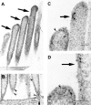
Immunoelectron microscopic localization of myosin Iβ in extrastriolar hair cells. Shown are the locations of side insertional plaques (A, C,D, arrows) and gold immunoreactivity (B–D) in medial extrastriolar Type B hair bundles. Note the presence of gold particles in the stereociliary rootlet (B, small arrow), ankle region (B, arrowheads), side plaques (C, D, arrowheads), and stereociliary tips (C, D,arrowheads). In D, there are 40 counted gold particles, but only three are indicated at the tipsof the arrowheads. As an example, thearrowhead on the top right of the panel is pointing to a single gold particle; in addition, there are two clearly identifiable particles immediately below this one. To theright of those two particles is a cluster that would not be included in the particle count because of the difficulty in discerning the number of particles present. Images were obtained at accelerating voltages of 60 kV (A–C) or 80 kV (D). Scale bars: 500 nm in A; 200 nm in B; 100 nm in C,D.
Fig. 6.
Fig. 6.
Immunoelectron microscopic localization of myosin Iβ in striolar hair cells. Shown are the locations of side insertional plaques (large arrows, A–C) and gold immunoreactivity (B, C) in stereocilia from an outer striolar Type C hair bundle. Note the presence of gold particles at the tip and side plaques (arrowheads, B, C). All images were obtained at an accelerating voltage of 60 kV. Scale bars: 500 nm in A; 100 nm in B,C.
Fig. 7.
Fig. 7.
Morphometric analysis of myosin Iβ localization, using coarse spatial bins. A, Schematic diagram of rectangular sampling grid with typical location of tip and side insertional plaques. B, Percentage of stereocilia with tip plaques located in the indicated bins. The tip plaque was always located in the apical-most kinociliary bin. C, Mean number and SD of gold particles found in each 200 nm kinociliary bin.D, Percentage of stereocilia with side plaques located in the indicated bins. The location of the side plaque on the nonkinociliary side was variable. E, Mean number and SD of gold particles found in each 200 nm nonkinociliary bin.
Fig. 8.
Fig. 8.
Morphometric analysis of myosin Iβ localization, using fine spatial bins. A, Schematic diagram of 75 nm square sampling grid with typical location of tip and side insertional plaques. B, Mean number and SD of gold particles located in each bin on the kinociliary side. There was no significant difference in the number of gold particles between any two pairs of bins on the kinociliary side of the stereocilium (p > 0.10). C, Mean number and SD of gold particles located in each bin on the nonkinociliary side. On this side the number of gold particles in the bin containing the side plaque was significantly greater than that in basal or apical bins located >200 nm from the side plaque (p < 0.05).

References

    1. Assad JA, Corey DP. An active motor model for adaptation by vertebrate hair cells. J Neurosci. 1992;12:3291–3309. - PMC - PubMed
    1. Assad JA, Shepherd GMG, Corey DP. Tip-link integrity and mechanical transduction in vertebrate hair cells. Neuron. 1991;7:985–994. - PubMed
    1. Baird RA. Morphological and electrophysiological properties of hair cells in the bullfrog utriculus. Ann NY Acad Sci. 1992;656:12–26. - PubMed
    1. Baird RA. Comparative transduction mechanisms of hair cells in the bullfrog utriculus. I. Responses to intracellular current. J Neurophysiol. 1994a;71:666–684. - PubMed
    1. Baird RA. Comparative transduction mechanisms of hair cells in the bullfrog utriculus. II. Sensitivity and response dynamics to hair bundle displacement. J Neurophysiol. 1994b;71:685–705. - PubMed

Publication types

LinkOut - more resources