

Aversive and appetitive events evoke the release of corticotropin-releasing hormone and bombesin-like peptides at the central nucleus of the amygdala
- PMID: 9614249
- PMCID: PMC6792703
- DOI: 10.1523/JNEUROSCI.18-12-04758.1998
Aversive and appetitive events evoke the release of corticotropin-releasing hormone and bombesin-like peptides at the central nucleus of the amygdala
Abstract
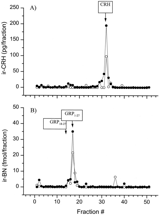
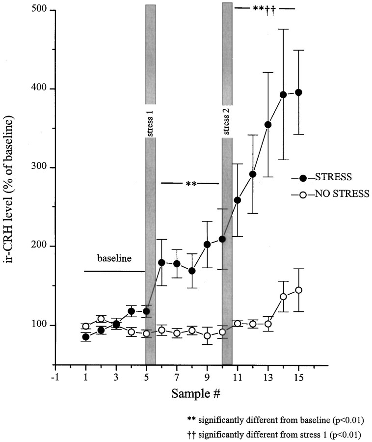
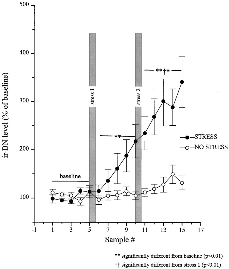
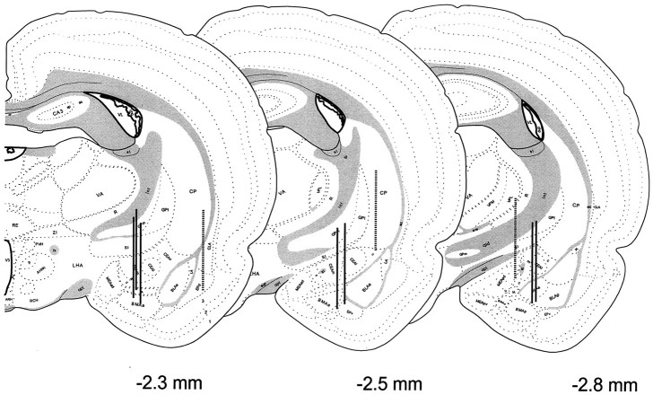
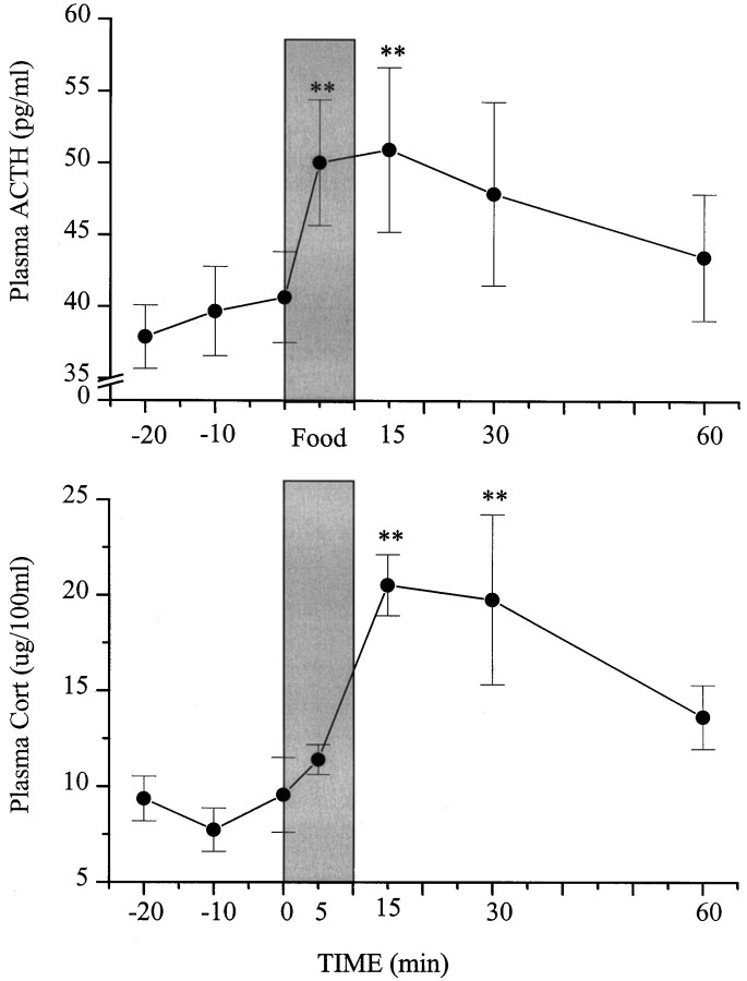
There is wide agreement that corticotropin-releasing hormone (CRH) systems within the brain are activated by stressful stimuli. There is also mounting evidence for the role of bombesin (BN)-like peptides in the mediation of the stress response. To date, however, the extent to which other stimuli increase the activity of these peptidergic systems has received little attention. In the present investigation we validated and used in vivo microdialysis sampling followed by ex vivo radioimmunoassays to monitor the release of CRH and BN-like peptides during appetitive (food intake) and stressful (restraint) events. It is demonstrated for the first time that the in vivo release of CRH and BN-like peptides at the central nucleus of the amygdala was markedly increased by both stressor exposure and food ingestion. In fact, the meal-elicited rise of CRH release was as great as that associated with 20 min of restraint stress. Paralleling these findings, circulating ACTH and corticosterone levels were also increased in response to both food intake and restraint. Contrary to the current views, these results indicate that either food ingestion is interpreted as a "stressful" event by certain neural circuits involving the central amygdala or that the CRH- and BN-related peptidergic systems may serve a much broader role than previously envisioned. Rather than evoking feelings of fear and anxiety, these systems may serve to draw attention to events or cues of biological significance, such as those associated with food availability as well as those posing a threat to survival.
Figures

Similar articles
-
Differential impact of predator or immobilization stressors on central corticotropin-releasing hormone and bombesin-like peptides in Fast and Slow seizing rat.Brain Res. 2001 Jul 6;906(1-2):60-73. doi: 10.1016/s0006-8993(01)02556-2. Brain Res. 2001. PMID: 11430862
-
Does amygdaloid corticotropin-releasing hormone (CRH) mediate anxiety-like behaviors? Dissociation of anxiogenic effects and CRH release.Eur J Neurosci. 2004 Jul;20(1):229-39. doi: 10.1111/j.1460-9568.2004.03468.x. Eur J Neurosci. 2004. PMID: 15245495
-
Central bombesin activates the hypothalamic-pituitary-adrenal axis. Effects on regional levels and release of corticotropin-releasing hormone and arginine-vasopressin.Neuroendocrinology. 2001 Mar;73(3):203-14. doi: 10.1159/000054637. Neuroendocrinology. 2001. PMID: 11307039
-
Differential involvement of amygdaloid CRH system(s) in the salience and valence of the stimuli.Prog Neuropsychopharmacol Biol Psychiatry. 2003 Dec;27(8):1201-12. doi: 10.1016/j.pnpbp.2003.09.014. Prog Neuropsychopharmacol Biol Psychiatry. 2003. PMID: 14659475 Review.
-
Role of bombesin-related peptides in the mediation or integration of the stress response.Cell Mol Life Sci. 2002 Feb;59(2):272-87. doi: 10.1007/s00018-002-8422-x. Cell Mol Life Sci. 2002. PMID: 11915944 Free PMC article. Review.
Cited by
-
The role of gastrin-releasing peptide on conditioned fear: differential cortical and amygdaloid responses in the rat.Psychopharmacology (Berl). 2006 Dec;189(3):287-96. doi: 10.1007/s00213-006-0585-y. Epub 2006 Oct 11. Psychopharmacology (Berl). 2006. PMID: 17033843
-
Social allostasis: anticipatory regulation of the internal milieu.Front Evol Neurosci. 2011 Jan 31;2:111. doi: 10.3389/fnevo.2010.00111. eCollection 2011. Front Evol Neurosci. 2011. PMID: 21369352 Free PMC article.
-
Noise stress changes mRNA expressions of corticotropin-releasing hormone, its receptors in amygdala, and anxiety-related behaviors.Noise Health. 2015 May-Jun;17(76):141-7. doi: 10.4103/1463-1741.155838. Noise Health. 2015. PMID: 25913553 Free PMC article.
-
Bombesin receptors as a novel anti-anxiety therapeutic target: BB1 receptor actions on anxiety through alterations of serotonin activity.J Neurosci. 2006 Oct 11;26(41):10387-96. doi: 10.1523/JNEUROSCI.1219-06.2006. J Neurosci. 2006. PMID: 17035523 Free PMC article.
-
Serotonergic responses to stress are enhanced in the central amygdala and inhibited in the ventral hippocampus during amphetamine withdrawal.Eur J Neurosci. 2014 Dec;40(11):3684-92. doi: 10.1111/ejn.12735. Epub 2014 Sep 19. Eur J Neurosci. 2014. PMID: 25234335 Free PMC article.
References
-
- Al-Damluji S, Iverson T, Thomas JM, Pendlebury DJ, Rees LH, Besser GM. Food-induced cortisol secretion is mediated by central alpha-1 adrenoceptor modulation of pituitary ACTH secretion. Clin Endocrinol. 1987;26:629–636. - PubMed
-
- Bartanusz V, Jezova D, Bertini LT, Tilders FJ, Aubry J-M, Kiss JZ. Stress-induced increase in vasopressin and corticotropin-releasing factor expression in hypophysiotrophic paraventricular neurons. Endocrinology. 1993;132:895–902. - PubMed
-
- Dallman MF, Akana SF, Strack AM, Hanson S, Sebastian RJ. The neural network that regulates energy balance is responsive to glucocorticoids and insulin and also regulates HPA axis responsivity at sites proximal to CRF neurons. Ann NY Acad Sci. 1995;771:730–742. - PubMed
-
- Davis M, Rainnie D, Cassell M. Neurotransmission in the rat amygdala related to fear and anxiety. Trends Neurosci. 1994;17:208–214. - PubMed
-
- Dunn AJ, Berridge CW. Physiological and behavioral responses to corticotropin-releasing factor administration: is CRF a mediator of anxiety or stress responses? Brain Res Rev. 1990;15:71–100. - PubMed
Publication types
MeSH terms
Substances
LinkOut - more resources
Full Text Sources
Other Literature Sources