

Aging alters the rhythmic expression of vasoactive intestinal polypeptide mRNA but not arginine vasopressin mRNA in the suprachiasmatic nuclei of female rats
- PMID: 9614250
- PMCID: PMC6792686
- DOI: 10.1523/JNEUROSCI.18-12-04767.1998
Aging alters the rhythmic expression of vasoactive intestinal polypeptide mRNA but not arginine vasopressin mRNA in the suprachiasmatic nuclei of female rats
Abstract
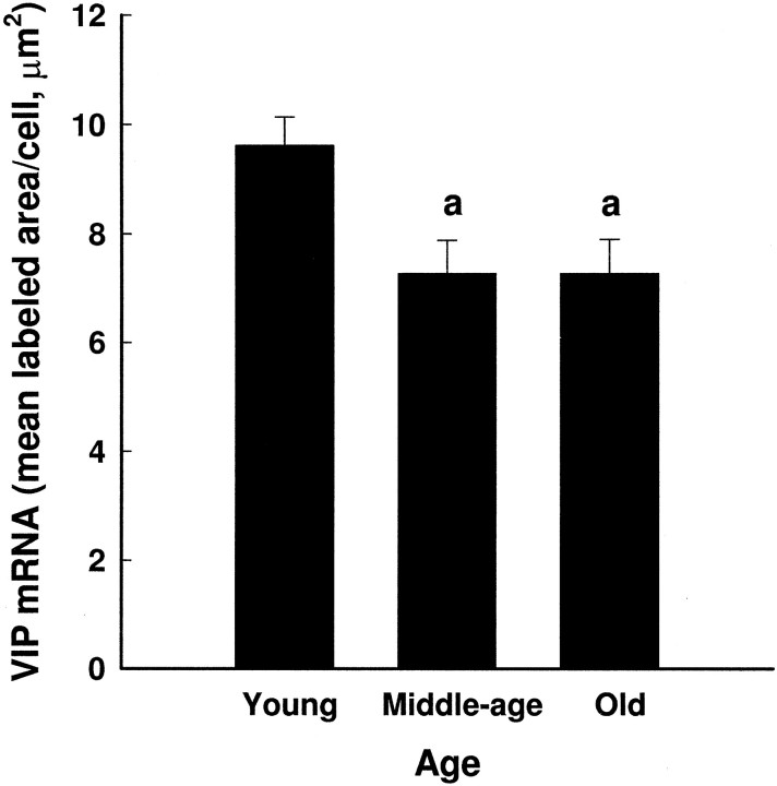
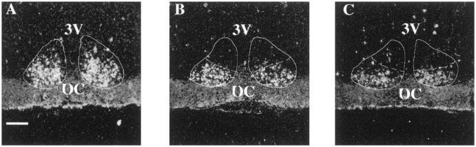
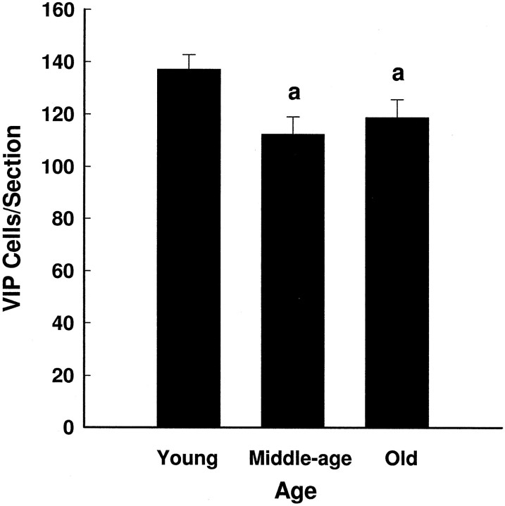
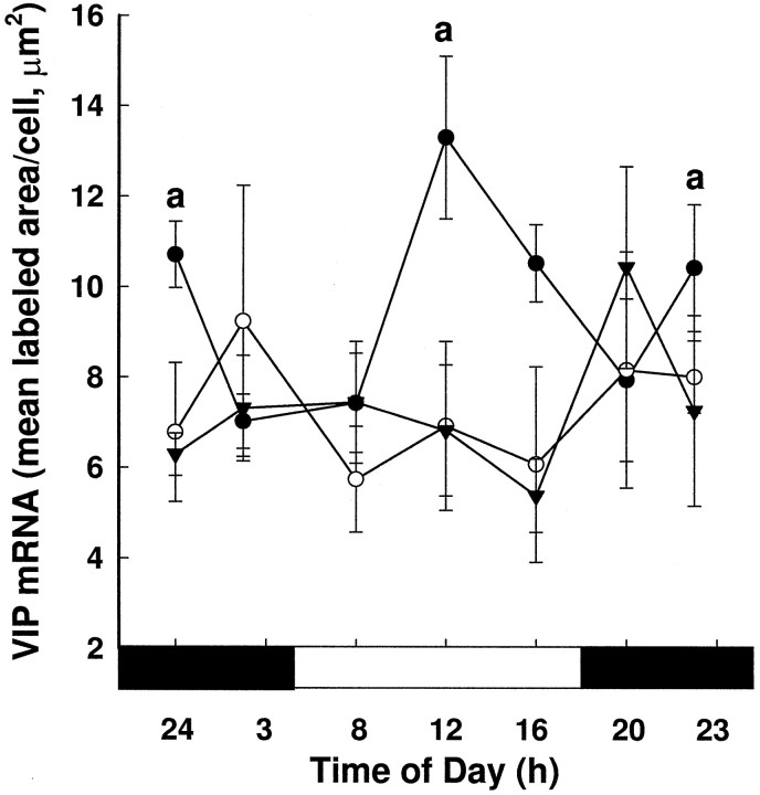
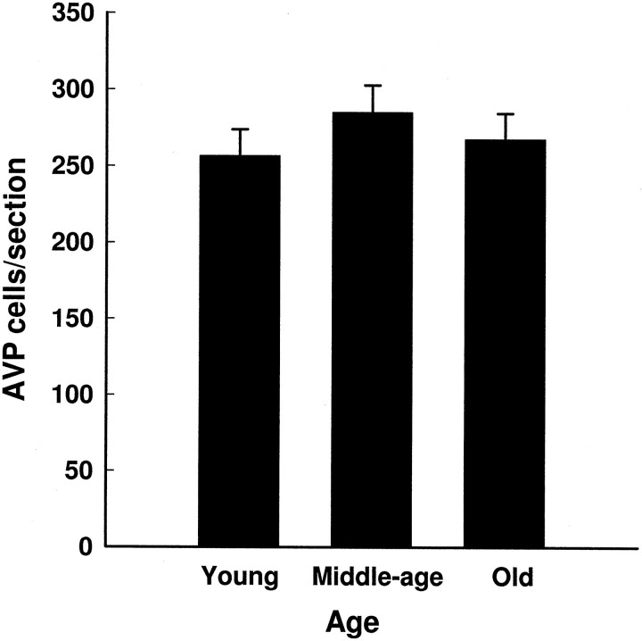
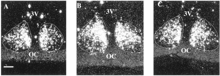
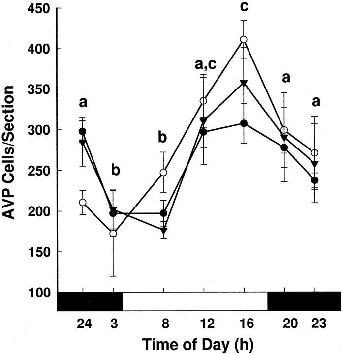
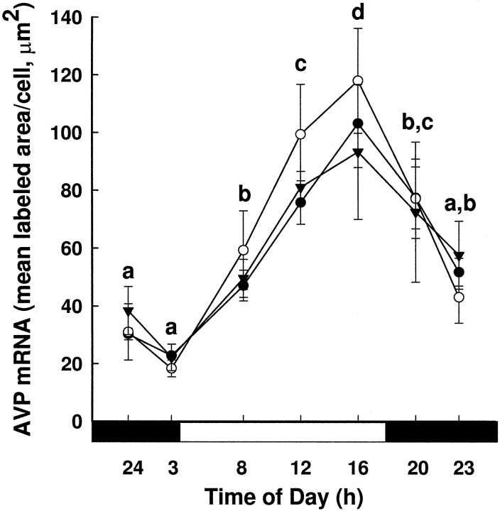
Our laboratory has shown that the ability of the suprachiasmatic nuclei (SCN) to regulate a number of rhythmic processes may be compromised by the time females reach middle age. Therefore, we examined the effects of aging on the rhythmic expression of two neuropeptides synthesized in the SCN, vasoactive intestinal polypeptide (VIP) and arginine vasopressin (AVP), using in situ hybridization. Because both VIP and AVP are outputs of the SCN, we hypothesized that age-related changes in rhythmicity are associated with alterations in the patterns of expression of these peptides. We found that VIP mRNA levels exhibited a 24 hr rhythm in young females, but by the time animals were middle-aged, this rhythm was gone. The attenuation of rhythmicity was associated with a decline in the level of mRNA per cell and in the number of cells in the SCN producing detectable VIP mRNA. AVP mRNA also showed a robust 24 hr rhythm in young females. However, in contrast to VIP, the AVP rhythm was not altered in the aging animals. The amount of mRNA per cell and the number of cells expressing AVP mRNA also was not affected with age. Based on these results we conclude that (1) various components of the SCN are differentially affected by aging; and (2) age-related changes in various rhythms may be attributable to changes in the ability of the SCN to transmit timing information to target sites. This may explain why the deterioration of various rhythmic processes occurs at different rates and at different times during the aging process.
Figures
Similar articles
-
Sex differences in the daily rhythm of vasoactive intestinal polypeptide but not arginine vasopressin messenger ribonucleic acid in the suprachiasmatic nuclei.Endocrinology. 1998 Oct;139(10):4189-96. doi: 10.1210/endo.139.10.6259. Endocrinology. 1998. PMID: 9751499
-
Changes in vasoactive intestinal peptide and arginine vasopressin expression in the suprachiasmatic nucleus of the rat brain following footshock stress.Neurosci Lett. 2007 Sep 25;425(2):99-104. doi: 10.1016/j.neulet.2007.08.044. Epub 2007 Aug 25. Neurosci Lett. 2007. PMID: 17826907 Free PMC article.
-
Aging selectively suppresses vasoactive intestinal peptide messenger RNA expression in the suprachiasmatic nucleus of the Syrian hamster.Brain Res Mol Brain Res. 2001 Mar 5;87(2):196-203. doi: 10.1016/s0169-328x(01)00015-8. Brain Res Mol Brain Res. 2001. PMID: 11245922
-
Vasopressin in circadian function of SCN.J Biosci. 2020;45:140. J Biosci. 2020. PMID: 33361631 Review.
-
An essential role for peptidergic signalling in the control of circadian rhythms in the suprachiasmatic nuclei.J Neuroendocrinol. 2003 Apr;15(4):335-8. doi: 10.1046/j.1365-2826.2003.01005.x. J Neuroendocrinol. 2003. PMID: 12622830 Review.
Cited by
-
Developmental alcohol and circadian clock function.Alcohol Res Health. 2001;25(2):136-40. Alcohol Res Health. 2001. PMID: 11584552 Free PMC article. Review.
-
New insights into the control of pulsatile GnRH release: the role of Kiss1/neurokinin B neurons.Front Endocrinol (Lausanne). 2012 Apr 2;3:48. doi: 10.3389/fendo.2012.00048. eCollection 2012. Front Endocrinol (Lausanne). 2012. PMID: 22649420 Free PMC article.
-
Proximate mechanisms driving circadian control of neuroendocrine function: Lessons from the young and old.Integr Comp Biol. 2009 Nov;49(5):519-37. doi: 10.1093/icb/icp041. Epub 2009 Jun 14. Integr Comp Biol. 2009. PMID: 21665838 Free PMC article.
-
Age-related decline of autocrine pituitary adenylate cyclase-activating polypeptide impairs angiogenic capacity of rat cerebromicrovascular endothelial cells.J Gerontol A Biol Sci Med Sci. 2015 Jun;70(6):665-74. doi: 10.1093/gerona/glu116. Epub 2014 Aug 18. J Gerontol A Biol Sci Med Sci. 2015. PMID: 25136000 Free PMC article.
-
A multi-level assessment of the bidirectional relationship between aging and the circadian clock.J Neurochem. 2021 Apr;157(1):73-94. doi: 10.1111/jnc.15286. Epub 2021 Jan 23. J Neurochem. 2021. PMID: 33370457 Free PMC article. Review.
References
-
- Albers HE, Gerall AA, Axelson JF. Effect of reproductive state on circadian periodicity of rat. Physiol Behav. 1981;26:21–25. - PubMed
-
- Albers HE, Stopa EG, Zoeller RT, Kauer JS, King JC, Fink JS, Mobtaker H, Wolfe H. Day-night variation in prepro-vasoactive intestinal peptide/peptide histidine isoleucine mRNA within the rat suprachiasmatic nucleus. Mol Brain Res. 1990;7:85–89. - PubMed
-
- Andreose JS, Fumagalli G, Clementi F. On the effect of aging on the distribution of vasoactive intestinal polypeptide and calcitonin gene-related peptide in the rat brain. Neurosci Lett. 1994;171:167–171. - PubMed
-
- Bosler O, Beaudet A. VIP neurons as prime synaptic targets for serotonin afferents in rat suprachiasmatic nucleus: a combined autoradiographic and immunocytochemical study. J Neurocytol. 1985;14:749–763. - PubMed
Publication types
MeSH terms
Substances
Grants and funding
LinkOut - more resources
Full Text Sources
Medical
Miscellaneous