. 1998 Jun;18(6):800-12.
doi: 10.1165/ajrcmb.18.6.3063.

Lung lymphocytes proliferate minimally in the murine pulmonary immune response to intratracheal sheep erythrocytes

Affiliations

Lung lymphocytes proliferate minimally in the murine pulmonary immune response to intratracheal sheep erythrocytes

G D Seitzman et al. Am J Respir Cell Mol Biol. 1998 Jun.

Abstract

The importance of in situ lymphocyte proliferation for net accumulation of lung lymphocytes during pulmonary immune responses and in immunologic lung diseases remains uncertain. Accordingly, we studied the experimental pulmonary immune response of antigen-primed C57BL/6 mice to intratracheal challenge with the particulate antigen sheep red blood cells. Uptake of nucleotide analogs (bromodeoxyuridine in vivo and tritiated thymidine in vitro), expression of the cell activation antigens CD25 and CD69 by flow cytometry, and response to the antimitotic agent hydroxyurea (in vivo) were measured. Although many lung lymphocytes and CD4+ T cells were CD25+ and CD69+, indicating recent activation, all techniques demonstrated that lung lymphocytes proliferated minimally in vivo. Blockade of cell division by hydroxyurea administration for 24 h did not significantly decrease lung lymphocyte accumulation on Day 3 after challenge. Lung lymphocytes also proliferated minimally in vitro (even on macrophage removal and despite addition of exogenous interleukin [IL]-2 or IL-4). However, lung lymphocytes responded vigorously to mitogens (immobilized anti-CD3, phytohemagglutinin, or concanavalin A), excluding global unresponsiveness to restimulation. Thus, in this model of pulmonary immunity, accumulation of lung lymphocytes does not require local T-cell proliferation and presumably depends instead on recruitment.


Figures

Figure 1
Figure 1
Flow cytometric analysis of in vivo lymphocyte proliferation. SRBC-primed C57BL/6 mice were challenged intratracheally with SRBC to induce a pulmonary immune response. On various days after challenge, BrdU (4 mgs/mouse IP) was administered in 1-3 doses at 20 minute intervals before the mouse was killed and cells harvested by BAL and dissection. Cells were stained for incorporated BrdU as described in Methods and were analyzed by flow cytometry. A-C. Representative histograms of cells within light scatter-defined gates from (A) bone marrow, (B) PTN, and (C) BAL. Data are from a single mouse four days after intratracheal antigen challenge. D. Kinetics of BrdU incorporation. Bars represent BrdU+ cells (defined by light scatter gating) in bone marrow (light cross hatching); paratracheal nodes (black); BAL (dark cross-hatching); and lung mince (light stippling). Data are mean ± SEM of 3 experiments, each consisting of pooled samples from 2-5 mice (except for bone marrows, which were assayed individually). *, significantly different from same tissue at other time-points, p <0.05, ANOVA with Fisher's PLSD post hoc testing.
Figure 2
Figure 2
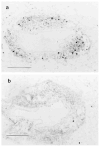
In situ demonstration of lung mononuclear cell proliferation. SRBC-primed C57BL/6 mice were challenged intratracheally with SRBC to induce a pulmonary immune response. BrdU (4 mgs/mouse) was administered IP in 1-3 doses at 20 minute intervals before the mouse was killed; lungs were processed histologically and sections were stained for BrdU incorporation by the immunogold technique as described in Methods. Representative serial photomicrographs of a single mouse four days after challenge. A. BrdU staining of individual cells within a perivenular inflammatory cell cuff. B. Control (irrelevant primary antibody). Similar results were obtained in three independent experiments. Scale indicates 100 μm.
Figure 3
Figure 3
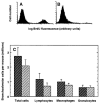
Effect of hydroxyurea treatment on lymphocyte accumulation in the lungs. SRBC-primed C57BL/6 mice were challenged intratracheally with SRBC to induce a pulmonary immune response. Two days later, hydroxyurea (1 mg/g body weight) or saline was administered IP for 24 hours (three separate injections at eight hour intervals). Mice were killed humanely 20 minutes after the last injection and cells were harvested from the lungs by BAL and from the bone marrow. A, B. Representative histograms of bone marrow from individual mice treated with saline (A) or hydroxyurea (B), showing effective systemic ablation of dividing cells in hydroxyurea-treated mice. C. Total and differential cell recovery in BAL. Control (saline injected) mice are represented by dark cross-hatching and hydroxyurea-treated mice by light cross-hatching. Data are mean ± SEM of ten mice per group assayed in two separate experiments. There are no significant differences between saline and hydroxyurea-treated groups, p >0.05, unpaired t test.
Figure 4
Figure 4
Intratracheal challenge results in CD25 expression by lung T cells. Peripheral blood lymphocytes (PBL) and BAL cells were obtained at various times after intratracheal challenge of SRBC-primed C57BL/6 mice. Cells were stained with pairs of conjugated monoclonal antibodies and analyzed by four parameter flow cytometry with light scatter gating to identify lymphocytes. A, B. Representative fluorescence data of (A) PBL and (B) BAL lymphocytes (defined by light-scatter gating). Data are depicted as 10% probability contour plots and represent cells from a single mouse. Horizontal axis is CD4 fluorescence and vertical axis is CD25 fluorescence; both axes are in arbitrary units on a four-decade logarithmic scale. The crossbars indicate the cut-offs for integration to determine double positive cells (upper right quadrant) as a percentage of all CD4+ T cells (upper right plus lower right quadrants). C, D. Time-course of CD25 expression by (C) total lymphocytes and (D) CD4+ T cells in lung and peripheral blood. Squares, BAL; circles, PBL. Open symbols denote results from normal mice. Data are mean ± SEM of 3-10 mice per time-point assayed individually in at least three separate experiments per time-point, except for normals and day zero, where lavage groups of 8-12 mice were pooled in each of four separate experiments. *, BAL sample significantly different from corresponding PBL sample, p < 0.05, unpaired t test.
Figure 5
Figure 5
Expression of the early activation antigen CD69 by BAL T cells. Splenic lymphocytes (PBL) and BAL cells were obtained at various times after IT challenge of SRBC primed C57BL/6 mice. Cells were stained with pairs of conjugated monoclonal antibodies and analyzed by four-parameter flow cytometry with light-scatter gating to identify lymphocytes. Representative fluorescence data of (A) splenic lymphocytes and (B) BAL lymphocytes (defined by light-scatter gating). Data are depicted as 10% probability contour plots and represent cells from a single mouse. Horizontal axis is CD4 fluorescence and vertical axis is CD69 fluorescence; both axes are in arbitrary units on a four decade logarithmic scale. Crossbars indicate the cutoffs for integration to determine CD69+ cells (upper right quadrant) as a percentage of all CD4+ T cells (upper right plus lower right quadrants). CD69 expression by (C) total lymphocytes and (D) CD4+ T cells in spleen ( ) and BAL ( ). N.D., not determined. Data are means 6 SEM of three mice per time point assayed individually in two to three separate experiments per time point. * BAL sample significantly different from corresponding splenic sample, P , 0.05, unpaired t test.
Figure 6
Figure 6
Spontaneous in vitro proliferation of lung lymphocytes from antigen-challenged mice. SRBC-primed C57BL/6 mice were challenged intratracheally with SRBC to induce a pulmonary immune response. On various days after challenge, cells obtained from BAL (dark cross-hatching), lung mince, (stippled), and lung mince post G-10 column (light cross-hatching) were placed in culture, pulsed immediately with 3H-Thd, and harvested 12-16 hours later. Values are expressed as mean ± SEM of at least three independent experiments at each time-point, each with replicates of 5-6 wells. *, significantly different from all other days within same cell-sample population; †, BAL significantly different from lung mince; ‡, BAL significantly different from post G-10 lung mince; § = lung mince significantly different from post G-10 lung mince; p < 0.05, ANOVA with Fisher's PLSD post hoc testing.
Figure 7
Figure 7
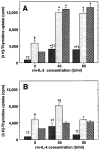
Effect of exogenous cytokines on lung lymphocyte proliferation in vitro. BAL (dark cross-hatching), lung mince (stippling), and lung mince post G-10 (light cross-hatching) were incubated for 24 hours with various concentrations of exogenous cytokines, then were pulsed with 3H-Thd and harvested after an additional 16-18 hour incubation. A. Incubation with rmIL-2. Data are mean ± standard error of 5-6 wells in a single experiment on cells obtained four days after intratracheal challenge, which is representative of three individual experiments. Similar results were obtained in each experiment using cells recovered at two and three days after intratracheal antigen challenge. B. Incubation with rmIL-4. Representative data of cells obtained at individual time-point post IT. Data are mean ± standard error of 5-6 wells in a single experiment on cells obtained four days after intratracheal challenge, which is representative of two individual experiments. *, significantly different from no cytokine incubation within same cell-sample population; p < 0.05, ANOVA with Dunnett's (two-tailed) post hoc testing; †, BAL significantly different from lung mince; ‡, BAL significantly different from post G-10 lung mince; § = lung mince significantly different from post G-10 lung mince; p < 0.05, ANOVA with Fisher's PLSD post hoc testing.
Figure 8
Figure 8
Proliferative response of lung lymphocytes to Con-A stimulation in vitro. BAL and lung mince mononuclear cells were harvested from SRBC-primed C57BL/6 mice four days after induction of a pulmonary immune response by IT challenge. Aliquots were depleted of phagocytic cells including macrophages by Sephadex G10 column. Cells were cultured for 2 days (light stippled bars), 3 days (cross-hatched bars), or 4 days (black bars) at 2 × 10⁵ cells per well in flat-bottomed 96-well plates in complete medium in the absence or presence of Con-A 10 μg/mL (an optimal dose). Lymphocyte proliferation was assessed by uptake of 3H-Thd during the final 16 hours of culture. Splenocytes in the same experiment gave 18,000-98,000 cpm. *, significantly different compared to corresponding condition without Con-A stimulation, p < 0.05, unpaired t test. Similar results were obtained in two separate experiments.
Figure 9
Figure 9
Proliferative response of lung lymphocytes to anti-CD3 stimulation in vitro. BAL and lung mince mononuclear cells were harvested from SRBC-primed C57BL/6 mice four days after induction of a pulmonary immune response by IT challenge. Aliquots were depleted of phagocytic cells including macrophages by Sephadex G10 column. Cells were cultured for 3 days (light stippled bars), 4 days (cross-hatched bars), or 5 days (black bars) at 2 × 10⁵ cells per well in flat-bottomed 96-well plates in complete medium in the absence or presence of immobilized hamster anti-murine CD3 (clone 145-2C-11). Lymphocyte proliferation was assessed by uptake of tritiated thymidine during the final 16 hours of culture. *, significantly different compared to corresponding condition without anti-CD3 stimulation, p < 0.05, unpaired t test. Note difference in scale from Figure 7. Similar results were obtained in two separate experiments.

Similar articles

Cited by

References

    1. Berman JS, Beer DJ, Theodore AC, Kornfeld H, Bernardo J, Center DM. Lymphocyte recruitment to the lung. Am Rev Respir Dis. 1990;142:238–257. - PubMed
    1. Sertl K, Takemura T, Tshachler E, Ferrans VJ, Kaliner MA, Shevach EM. Dendritic cells with antigen-presenting capability reside in airway epithelium, lung parenchyma and visceral pleura. J Exp Med. 1986;163:436–451. - PMC - PubMed
    1. Sitrin RG, Ansfield MJ, Kaltreider HB. The effect of pulmonary surface-active material on the generation and expression of murine B- and T- lymphocyte effector functions in vitro. Exp Lung Res. 1985;9:85–97. - PubMed
    1. Roth MD, Golub SH. Human pulmonary macrophages utilize prostaglandins and transforming growth factor β1 to suppress lymphocyte activation. J Leukocyte Biol. 1993;53:366–371. - PubMed
    1. Paine R, III, Chavis A, Gaposchkin D, Christensen P, Mody CH, Turka LA, Toews GB. A factor secreted by a human pulmonary alveolar epithelial-like cell line blocks T-cell proliferation between G1 and S phase. Am J Respir Cell Mol Biol. 1992;6:658–666. - PubMed

Publication types

MeSH terms