Review
. 1998 Jun;62(2):275-93.
doi: 10.1128/MMBR.62.2.275-293.1998.

Short-sequence DNA repeats in prokaryotic genomes

Affiliations
Review

Short-sequence DNA repeats in prokaryotic genomes

A van Belkum et al. Microbiol Mol Biol Rev. 1998 Jun.

Abstract

Short-sequence DNA repeat (SSR) loci can be identified in all eukaryotic and many prokaryotic genomes. These loci harbor short or long stretches of repeated nucleotide sequence motifs. DNA sequence motifs in a single locus can be identical and/or heterogeneous. SSRs are encountered in many different branches of the prokaryote kingdom. They are found in genes encoding products as diverse as microbial surface components recognizing adhesive matrix molecules and specific bacterial virulence factors such as lipopolysaccharide-modifying enzymes or adhesins. SSRs enable genetic and consequently phenotypic flexibility. SSRs function at various levels of gene expression regulation. Variations in the number of repeat units per locus or changes in the nature of the individual repeat sequences may result from recombination processes or polymerase inadequacy such as slipped-strand mispairing (SSM), either alone or in combination with DNA repair deficiencies. These rather complex phenomena can occur with relative ease, with SSM approaching a frequency of 10(-4) per bacterial cell division and allowing high-frequency genetic switching. Bacteria use this random strategy to adapt their genetic repertoire in response to selective environmental pressure. SSR-mediated variation has important implications for bacterial pathogenesis and evolutionary fitness. Molecular analysis of changes in SSRs allows epidemiological studies on the spread of pathogenic bacteria. The occurrence, evolution and function of SSRs, and the molecular methods used to analyze them are discussed in the context of responsiveness to environmental factors, bacterial pathogenicity, epidemiology, and the availability of full-genome sequences for increasing numbers of microorganisms, especially those that are medically relevant.


Figures

FIG. 1
FIG. 1
Schematic survey of SSRs. (A) Examples of homogeneous simple sequence motifs consisting of repeat units varying from 1 (homopolymeric tract) to 6 nucleotides in length. (B) Example of a composite, heterogeneous repeat built from three 3-nucleotide units, two 5-nucleotide units, and seven 2-nucleotide motifs. (C) Comparative analysis of four different repeats built from three 10-nucleotide units showing degeneracy among units. Identity of the nucleotide sequences B through D with the consensus given in sequence A is indicated by dashes.
FIG. 2
FIG. 2
Schematic representation of the mechanism of SSM during replication, which results in shortening or lengthening of SSRs. Individual repeat units are identified by arrows; bulging is the presence of non-base-pair base residues interrupting a regular 2-strand DNA helix. Bulging in the nascent strand leads to a larger number of repeat units; bulging in the template strand results in a smaller numbers of units. During replication, bulges can occur in both strands, and the effect of insertion or deletion can be neutralized by occurrence of the adverse event. The number of repeat units can decrease or increase by multiple repeats once multiple bulging in one strand has occurred.
FIG. 3
FIG. 3
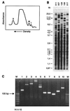
Molecular identification of SSR-type DNA. (A) Microdensitometer tracing of human leukocyte native DNA centrifuged to equilibrium in density gradients with an analytical ultracentrifuge. The satellite peaks representing repetitive DNA fractions displaying aberrant densities are indicated (I to III). (B) DNA fingerprints of human individuals generated by probing with repetitive DNA. DNA from five individuals (identified by numerals above the lanes) was digested with a restriction enzyme, the fragments were separated by electrophoresis, and after blotting, the resulting Southern blot was probed with a synthetic oligonucleotide SSR consisting of 10 units of a TTAGG motif. The autoradiograph shows that this SSR is widely dispersed throughout the human genome and clearly depicts the hypervariability in the observed banding patterns. (C) PCR amplification of SSR regions. A specific SSR was identified in the genome of H. influenzae, and primers bordering the repetitive motif were synthesized. When DNA from bacterial strains 1 to 10 was used as template, various amplicons were generated, most of them showing clear differences in length (related to the number of repeat units present). Lanes M contain 10-bp molecular size markers.
FIG. 4
FIG. 4
Hypothetical model for the mechanism of fimbrial phase variation in H. influenzae. Transcription of the two divergently oriented genes hifA and hifB is controlled by a variable sequence of reiterated TA units (long open box) in their combined promoter region. In nine units in this SSR, the putative −10 and −35 promoter sequences for both hifA (hatched boxes) and hifB (short stippled boxes) are separated by 14 bases, which does not allow transcription of either gene. With 10 or 11 TA units, the −10 and −35 motifs are separated by 16 or 18 bases, respectively, allowing transcription of both genes, resulting in the expression of fimbriae. A spacing of 16 bases results in the highest level of fimbriation. With 12 TA units, hifA and hifB transcription can take place with alternative −35 promoter sequences (narrow boxes), which are separated by 16 bases from the corresponding −10 sequences. Solid boxes represent the +1 transcriptional start point. Reprinted from reference with permission of the publisher.
FIG. 5
FIG. 5
Schematic models of MSCRAMMs from S. aureus. The MSCRAMMs shown are the ones with affinity for fibronectin (FnbpA and FnbpB), collagen (CNA), and fibrinogen (CLF, clumping factor). S, signal sequence; U or A, unique nonrepetitive sequence; δ, upstream repeat sequences; D, B, or R: repeated domains; W, cell wall-spanning domain; M, hydrophobic membrane-spanning domain; C, positively charged carboxy terminus. Reprinted from reference with permission of the authors and the publisher.
FIG. 6
FIG. 6
Epidemiology of H. influenzae infections. During an outbreak, strains 1 to 13 were isolated from different patients and compared to seven nonrelated clinical isolates (strains 14 to 20) on the basis of SSR polymorphisms. Four different SSRs were analyzed for the occurrence of length variability, and assays 3-1, 6-1, and 6-2 correctly identified the epidemic isolates as identical. The controls clearly differed in some instances. Interestingly, assay 5-2 also revealed major polymorphisms among the epidemic isolates, possibly identifying a “contingency locus” that is tailoring colonization or infection of a range of individual human hosts. Lanes M contain molecular size markers; the arrows on the right identify a 100-bp DNA fragment. Reprinted from reference with permission of the American Society for Microbiology.

References

    1. Aaltonen L A, Peltomaki P, Leach F S, Sistonen P, Pylkkanen L, Mecklin J, Jarvinen H, Powell S M, Jen J, Hamilton S R, Petersen G M, Kinzler K W, Vogelstein B, de la Chapelle A. Clues to the pathogenesis of familial colorectal cancer. Science. 1993;260:812–816. - PubMed
    1. Allred D R, McGuire T C, Palmer G H, Leib S R, Harkins T M, McElwain T F, Barbet A F. Molecular basis for surface antigen size polymorphisms and conservation of a neutralization sensitive epitope in Anaplasma marginale. Proc Natl Acad Sci USA. 1990;87:3220–3224. - PMC - PubMed
    1. Andersen G L, Simchock J M, Wilson K H. Identification of a region of variability among Bacillus anthracis strains and related species. J Bacteriol. 1996;178:377–384. - PMC - PubMed
    1. Arbeit R D. Laboratory procedures for the epidemiologic analysis of staphylococci. In: Archer G L, Grassley K B, editors. The biology of staphylococci. New York, N.Y: Churchill Livingstone; 1996. pp. 253–286.
    1. Asayama M, Kabasawa M, Takahashi I, Aida T, Shirai M. Highly repetitive sequences and characteristics of genomic DNA in unicellular cyanobacterial strains. FEMS Microbiol Lett. 1996;137:175–181. - PubMed

LinkOut - more resources