Review
. 1998 Jun;62(2):362-78.
doi: 10.1128/MMBR.62.2.362-378.1998.

Regulation of X-chromosome inactivation in development in mice and humans

Affiliations
Review

Regulation of X-chromosome inactivation in development in mice and humans

T Goto et al. Microbiol Mol Biol Rev. 1998 Jun.

Abstract

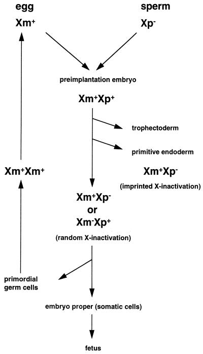
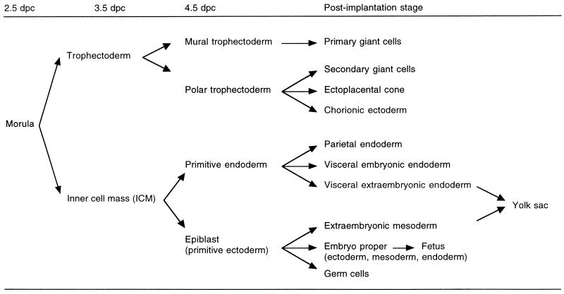
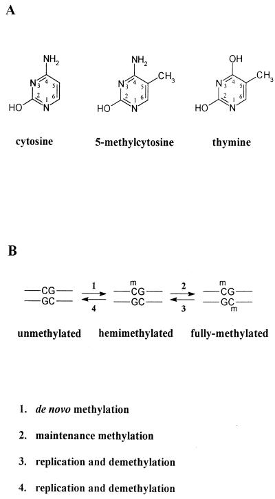
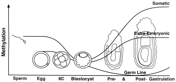
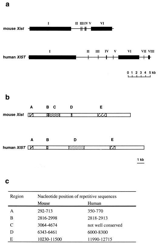
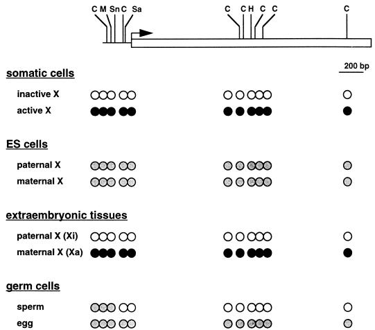
Dosage compensation for X-linked genes in mammals is accomplished by inactivating one of the two X chromosomes in females. X-chromosome inactivation (XCI) occurs during development, coupled with cell differentiation. In somatic cells, XCI is random, whereas in extraembryonic tissues, XCI is imprinted in that the paternally inherited X chromosome is preferentially inactivated. Inactivation is initiated from an X-linked locus, the X-inactivation center (Xic), and inactivity spreads along the chromosome toward both ends. XCI is established by complex mechanisms, including DNA methylation, heterochromatinization, and late replication. Once established, inactivity is stably maintained in subsequent cell generations. The function of an X-linked regulatory gene, Xist, is critically involved in XCI. The Xist gene maps to the Xic, it is transcribed only from the inactive X chromosome, and the Xist RNA associates with the inactive X chromosome in the nucleus. Investigations with Xist-containing transgenes and with deletions of the Xist gene have shown that the Xist gene is required in cis for XCI. Regulation of XCI is therefore accomplished through regulation of Xist. Transcription of the Xist gene is itself regulated by DNA methylation. Hence, the differential methylation of the Xist gene observed in sperm and eggs and its recognition by protein binding constitute the most likely mechanism regulating imprinted preferential expression of the paternal allele in preimplantation embryos and imprinted paternal XCI in extraembryonic tissues. This article reviews the mechanisms underlying XCI and recent advances elucidating the functions of the Xist gene in mice and humans.


Figures

FIG. 1
FIG. 1
Activity of X chromosomes in female mouse development. The maternally inherited X chromosome (Xm) in the egg is active, and the paternally inherited X chromosome (Xp) in the sperm is inactive. Soon after fertilization, Xp is reactivated and both Xm and Xp are active (Xm+Xp+) in preimplantation embryos. In the first delineating cell lineages, the trophectoderm and the primitive endoderm, which differentiate at 3.5 and 4.5 dpc, respectively, Xp is preferentially inactivated (Xm+Xp; imprinted X inactivation). In the epiblast cells, which will give rise to the germ cells and somatic cells of the embryo proper, either Xm or Xp is inactivated (Xm+Xp or XmXp+; random X inactivation). In the primordial germ cells, the inactive X chromosome is reactivated at around 12.5 dpc, when the female germ cells enter meiosis; hence, both X chromosomes are active (Xm+Xm+) throughout oogenesis. + denotes the active X chromosome, and  denotes the inactive X chromosome.
FIG. 2
FIG. 2
Diagrammatic representation of cell differentiation during mouse development. The trophectoderm is the first cell lineage, which differentiates at 3.5 dpc. Then the primitive endoderm differentiates, at 4.5 dpc. In the derivatives of these two extraembryonic cell lineages, XCI is nonrandom, in that the paternally inherited X chromosome is preferentially inactivated (see Table 1 for summary). In contrast, XCI is random in the epiblast, which gives rise to the yolk sac mesoderm, the embryo proper, and the primordial germ cells; i.e., either the paternal or the maternal X chromosome is inactivated in a given cell.
FIG. 3
FIG. 3
Methylation of cytosine residues in the mammalian genome. (A) Structure of the cytosine residue and its methylated derivative, 5-methylcytosine, which is methylated at the 5 position of the carbon ring of the cytosine residue. The structure of the thymine residue is also shown, to illustrate that mutational deamination at the 4 position of 5-methylcytosine results in thymine. (B) After replication, daughter strands of fully methylated DNA are hemimethylated (reaction 3) and the original pattern of DNA methylation is maintained by the DNA methyltransferase (reaction 2), which preferentially methylates the cytosine residues at hemimethylated CpG sites. Further replication without methylation of the hemimethylated DNA results in fully unmethylated DNA (reaction 4). De novo methylation (reaction 1) is also considered to be mediated by the DNA methyltransferase, although the efficiency of de novo methylation is low.
FIG. 4
FIG. 4
Changes in DNA methylation in early development. Sperm DNA is overall more methylated than egg DNA but less methylated than somatic DNA. During preimplantation development, gamete-specific patterns of methylation are erased by the genome-wide demethylation, tending toward a ground state of absence of methylation in the ICM of the blastocyst and in the primordial germ cells. Around the time of implantation and gastrulation, stage- and tissue-specific patterns of DNA methylation are created de novo. In the extraembryonic tissues, the methylation level is globally lower than that in somatic tissues. It is hypothesized that demethylation continues in the germ line until the onset of establishment of new egg- or sperm-specific patterns of methylation, including the differences in methylation between the gametes that will survive as imprints in the next generation. Imprinting can be viewed as differences in gametic DNA modification (differences between sperm and egg) that survive erasure during preimplantation development and thus are perpetuated into the soma. Reprinted from reference with permission of the publisher.
FIG. 5
FIG. 5
Structure of the Xist gene. (a) The mouse Xist gene and the human XIST gene comprise six and eight exons, respectively. (b) Schematic representation of the Xist RNA. The mouse Xist RNA is 15 kb long, and the human XIST RNA is 17 kb long. Corresponding conserved repetitive sequences in exons 1 (regions A, B, C, and D) and 6 (region E) are indicated by similarly hatched regions. The numbers of repeats are different between the mouse and human. (c) Nucleotide positions of the repetitive sequences are shown. Data are from references and .
FIG. 6
FIG. 6
DNA methylation of the Xist gene. The horizontal line indicates the Xist promoter region. The open rectangle indicates the 5′ portion of the exon 1. The hooked arrow denotes the transcription start site. The methylation-sensitive restriction enzyme sites so far tested are shown: C, HhaI (CfoI); M, MluI; Sn, SnaBI, Sa, SacII; H, HpaII. The methylation status of the methylation-sensitive restriction sites is shown. Open circles indicate lack of methylation; solid circles indicate complete methylation; shaded circles indicate mosaic methylation in that a particular site is methylated with a certain probability on each allele. It is hypothesized that the promoter and the 5′ end of exon 1 are mosaically methylated in eggs. Xi, inactive X chromosome; Xa, active X chromosome. Data for somatic cells and extraembryonic tissues are from reference ; data for ES cells are from references and ; and data for germ cells are from references , , , , and .

Similar articles

Cited by

References

    1. Adams R L P, Burdon R H, McKinnon K, Rinaldi A. Stimulation of de novo methylation following limited proteolysis of mouse ascites DNA methylase. FEBS Lett. 1983;163:194–198. - PubMed
    1. Adams R L P. Eukaryotic DNA methyltransferases—structure and function. Bioessays. 1995;17:139–145. - PubMed
    1. Antequera F, Boyes J, Bird A P. High levels of de novo methylation and altered chromatin structure at CpG islands in cell lines. Cell. 1990;62:503–514. - PubMed
    1. Ariel M, Robinson E, McCarrey J R, Cedar H. Gamete-specific methylation correlates with imprinting of the murine Xist gene. Nat Genet. 1995;9:312–315. - PubMed
    1. Barlow D P, Stoger R, Herrmann B G, Saito K, Schweiter N. The mouse insulin-like growth factor type-2 receptor is imprinted and closely linked to the Tme locus. Nature. 1991;349:84–87. - PubMed

Substances

LinkOut - more resources