. 1998 Jun 9;95(12):6791-6.
doi: 10.1073/pnas.95.12.6791.

p21(WAF1) is required for butyrate-mediated growth inhibition of human colon cancer cells

Affiliations

p21(WAF1) is required for butyrate-mediated growth inhibition of human colon cancer cells

S Y Archer et al. Proc Natl Acad Sci U S A. .

Abstract

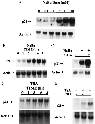
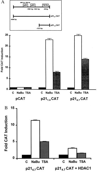
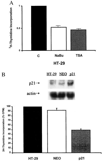
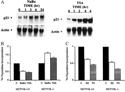
A diet high in fiber is associated with a decreased incidence and growth of colon cancers. Butyrate, a four-carbon short-chain fatty acid product of fiber fermentation within the colon, appears to mediate these salutary effects. We sought to determine the molecular mechanism by which butyrate mediates growth inhibition of colonic cancer cells and thereby to elucidate the molecular link between a high-fiber diet and the arrest of colon carcinogenesis. We show that concomitant with growth arrest, butyrate induces p21 mRNA expression in an immediate-early fashion, through transactivation of a promoter cis-element(s) located within 1.4 kb of the transcriptional start site, independent of p53 binding. Studies using the specific histone hyperacetylating agent, trichostatin A, and histone deacetylase 1 indicate that growth arrest and p21 induction occur through a mechanism involving histone hyperacetylation. We show the critical importance of p21 in butyrate-mediated growth arrest by first confirming that stable overexpression of the p21 gene is able to cause growth arrest in the human colon carcinoma cell line, HT-29. Furthermore, using p21-deleted HCT116 human colon carcinoma cells, we provide convincing evidence that p21 is required for growth arrest to occur in response to histone hyperacetylation, but not for serum starvation nor postconfluent growth. Thus, p21 appears to be a critical effector of butyrate-induced growth arrest in colonic cancer cells, and may be an important molecular link between a high-fiber diet and the prevention of colon carcinogenesis.


Figures

Figure 1
Figure 1
NaBu and TSA induce p21 mRNA expression in HT-29 cells in an immediate-early fashion. (A) Dose response for p21 induction by NaBu. HT-29 cells were treated with various concentration of NaBu, 0–20 mM for 24 hr, and p21 mRNA expression was examined by Northern blot analyses. (B) Time course for induction of p21 by NaBu. Cells were treated with 5 mM NaBu for varying lengths of time, from 0 to 24 hr, and p21 induction was examined by Northern analysis. (C) p21 induction by NaBu is not blocked by protein synthesis inhibition. HT-29 cells were treated concomitantly with 5 mM NaBu and the protein synthesis inhibitor, cycloheximide (CHX, 10 μg/ml), for 48 hr. (D) Time course for p21 induction by TSA. Cells were treated with 0.3 μM TSA for varying lengths of time. (E) p21 induction by TSA is not blocked by protein synthesis inhibition. Cells were treated concomitantly with 0.3 μM TSA and 10 μg/ml CHX for 6 hr. Representative Northern blots (n = 4) are depicted in each figure. The actin control is shown in the lower panel. Twenty micrograms of total RNA was loaded in each lane, with equal loading verified by ethidium bromide staining.
Figure 2
Figure 2
Overexpression of histone deacetylase (HDAC1) blocks induction of the p21 promoter by NaBu and TSA. (A) NaBu and TSA induce p21 promoter activity. HT-29 cells were transiently transfected with CAT reporter plasmids under the control of various amounts of the mouse p21 promoter (0–4.7 kb upstream from the transcriptional start site). Transfectants were treated without or with 5 mM NaBu or 0.3 μM TSA, and total cellular protein was examined for CAT expression. Results are shown for the 0-kb (pCAT), 1.4-kb (p211.4CAT), and 4.7-kb (p214.7CAT) p21 promoter constructs and are expressed as fold CAT induction compared with the untreated negative control, arbitrarily taken as 1. (B) Cotransfection of HDAC1 blocks p21 promoter induction by NaBu and TSA. Transfections were performed with the p214.7 CAT −/+ HDAC1, and cells were treated without or with 5 mM NaBu or 0.3 μM TSA. The results are expressed as fold CAT induction compared with the untreated negative control (arbitrarily taken as 1). All results are shown as mean ± SEM (n ≥ 4, in all cases).
Figure 3
Figure 3
NaBu, TSA, and p21 overexpression inhibit growth of HT-29 cells. (A) NaBu and TSA inhibit growth of HT-29 cells. Cells were plated at equal density in six-well plates. At 80% confluence, cells were treated without or with 5 mM NaBu or 0.3 μM TSA for 24 hr. 3H-thymidine (1 μCi/ml) was added for the last 6 hr of treatment, and incorporation was measured by scintillation counting. Treatment results are expressed as percent change compared with untreated negative control, arbitrarily taken as 1. (B) p21 overexpression inhibits growth of HT-29 cells. Cells were stably transfected with an expression plasmid containing the human p21 gene, and overexpression of the gene in pooled stable transfectants compared with the parental and NEO controls (n = 5 each) was determined by Northern analyses (see Inset). 3H-thymidine incorporation was measured in exponentially growing cells under basal conditions, and results are expressed as percent change compared with parental control, arbitrarily taken as 100%, and are shown as mean ± SEM (n ≥ 4, in all cases).
Figure 4
Figure 4
p21 is required for NaBu-induced growth inhibition of colon cancer cells. (A) p21 mRNA is induced early in HCT116 +/+ cells. Cells were treated at 80% confluence with 1 mM NaBu or 0.15 μM TSA (higher concentrations of either agent led to cell death), and p21 mRNA expression was examined by Northern analysis. (B) p21 deletion prevents growth inhibition by butyrate and TSA. HCT116 +/+ and −/− cells were treated with 1 mM NaBu or 0.15 μM TSA for 24 hr, and 1 μCi/ml 3H-thymidine was added for the last 6 hr of treatment. Incorporation was measured by scintillation counting. (C) HCT116 −/− cells are growth-inhibited by serum starvation and postconfluent growth. HCT116 +/+ and −/− cells were serum-starved for 24 hr or grown to 2 weeks postconfluence, and 3H-thymidine incorporation was measured. Cells grown in standard McCoy’s 5A medium with 10% fetal bovine serum and that were preconfluent were used as controls. Results are expressed as percent change compared with control groups (arbitrarily taken as 1) and are shown as mean ± SEM (n ≥ 4, in all cases).

Similar articles

Cited by

References

    1. Boring C C, Squires T S, Tong T, Montgomery S. CA Cancer J Clin. 1994;44:7–26. - PubMed
    1. Trock B, Lanza E, Greenwald P. J Natl Cancer Inst. 1990;82:650–661. - PubMed
    1. McIntyre A, Gibson P R, Young G P. Gut. 1993;34:386–391. - PMC - PubMed
    1. Barnard J A, Warwick G. Cell Growth Differ. 1993;4:495–501. - PubMed
    1. Whitehead R H, Young G P, Bhathal P S. Gut. 1986;27:1457–1463. - PMC - PubMed

Publication types

MeSH terms