. 1998 Jun 9;95(12):6887-92.
doi: 10.1073/pnas.95.12.6887.

Nucleolar localization of the Werner syndrome protein in human cells

Affiliations

Nucleolar localization of the Werner syndrome protein in human cells

R A Marciniak et al. Proc Natl Acad Sci U S A. .

Abstract

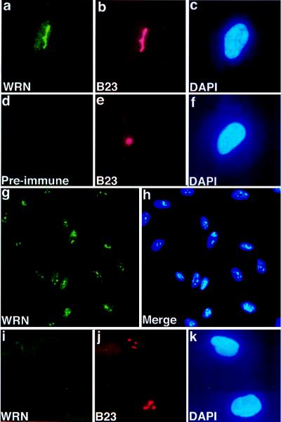
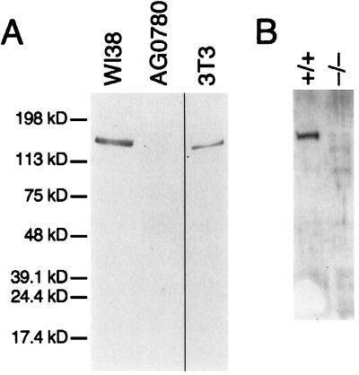
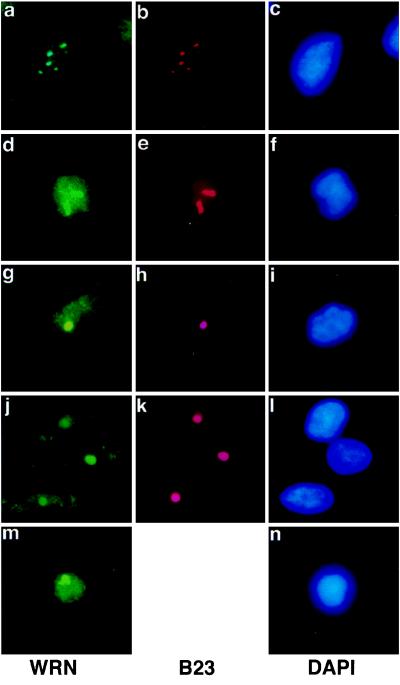
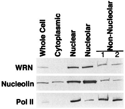
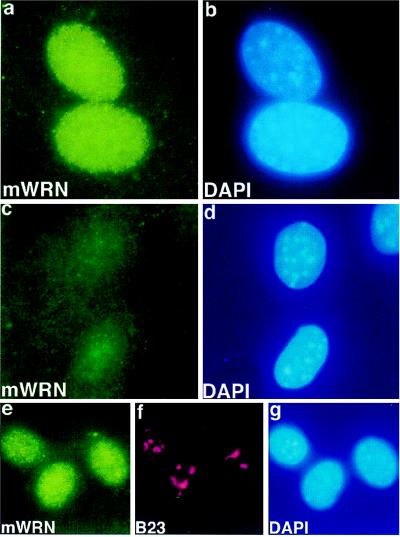
Werner Syndrome (WS) is a human genetic disorder with many features of premature aging. The gene defective in WS (WRN) has been cloned and encodes a protein homologous to several helicases, including Escherichia coli RecQ, the human Bloom syndrome protein (BLM), and Saccharomyces cerevisiae Sgs1p. To better define the function of WRN protein we have determined its subcellular localization. Indirect immunofluorescence using polyclonal anti-human WRN shows a predominant nucleolar localization. Studies of WRN mutant cells lines confirmed the specificity of antibody recognition. No difference was seen in the subcellular localization of the WRN protein in a variety of normal and transformed human cell lines, including both carcinomas and sarcomas. The nucleolar localization of human WRN protein was supported by the finding that upon biochemical subcellular fractionation, WRN protein is present in an increased concentration in a subnuclear fraction enriched for nucleolar proteins. We have also determined the subcellular localization of the mouse WRN homologue (mWRN). In contrast to human WRN protein, mWRN protein is present diffusely throughout the nucleus. Understanding the function of WRN in these organisms of vastly differing lifespan may yield new insights into the mechanisms of lifespan determination.


Figures

Figure 1
Figure 1
Nucleolar localization of WRN in human cell lines. Immunofluorescence images of human fetal lung fibroblasts (WI38, a–h) or adult skin fibroblasts derived from a patient with WS (cell line AG0780, i–k). Indirect immunofluorescence was performed on methanol/acetone-fixed cells, using anti-WRN antibody (a, d, g) or antibody to nucleolar protein B23 (b, e, j) or by DAPI, which binds to nuclear DNA (c, f, h, k). The bound antibody is visualized with a fluorescein-conjugated secondary antibody (green fluorescence), or Texas red-conjugated secondary antibody (red fluorescence). WI38 cells were treated consecutively with anti-WRN (a), anti-nucleolar protein B23 (b), or DAPI (c). No fluorescence was seen when preimmune serum was used in place of anti-WRN antibody (d) and cells were treated consecutively with anti-B23 (e) and DAPI (f). A low-power view of WI38 cells treated with anti-WRN shows little cell-to-cell variation in the immunofluorescence signal (g), as also shown in the composite photomicrograph of WI38 cells stained with anti-WRN and DAPI (h). Fibroblasts from a patient with WS show no staining when treated with anti-WRN (i), although no difference in gross nucleolar (j) or nuclear (k) morphology is observed. (g and h, × 200 ; all others, × 500.)
Figure 2
Figure 2
Immunoblot of human and mouse WRN proteins. Whole cell extracts of the indicated cell lines were prepared, and 10 μg of each was analyzed by Western blotting. (A) Immunoblot of 8% polyacrylamide gel, using anti-WRN antibody. Anti-human WRN recognizes a single protein of apparent mobility corresponding to 170 kDa in fibroblast cell line WI38. In a WRN mutant fibroblast cell line, AG0780, no reactive protein is seen. A single protein of slightly increased mobility is seen in an extract prepared from mouse 3T3 cells, in agreement with the previous identification of a single, highly conserved WRN homologue in mouse (38). (B) Immunoblot of a 7.5% polyacrylamide gel, using anti-mWRN antibody. Anti-mWRN recognizes a single protein of apparent mobility corresponding to 165 kDa in normal mouse ear fibroblasts (+/+). No cross-reacting protein is seen in ear fibroblasts prepared from a mouse in which mWRN has been disrupted (−/−).
Figure 3
Figure 3
WRN nucleolar localization in multiple cell lines. Cells were subjected to double immunolabeling with anti-WRN and anti-nucleolar protein B23, as described in the legend to Fig. 1. In each row, Left shows the anti-WRN signal, Center shows the anti-B23 (anti-nucleolar signal), and Right shows the DAPI (nuclear) signal. (a–c) WI38 75.1, simian virus 40-transformed human fetal lung fibroblast cell line. (d–f) HeLa, human cervical carcinoma cell line. (g–i) PC-3, prostatic adenocarcinoma. (j–l) GCT, fibrous histiocytoma. (m and n) AG07877, lymphoblastoid cell line. (×500.)
Figure 4
Figure 4
Distribution of WRN protein among HeLa subcellular fractions. Extracts of HeLa cells were prepared and fractionated into cytoplasmic, nuclear, nucleolar, and nonnucleolar fractions. A 10-μg sample of each fraction was separated by electrophoresis on an SDS/4–15% polyacrylamide gradient gel and then analyzed by Western blotting. Blots, prepared in parallel, were probed with the antibodies indicated to the left. To control for the fidelity of the fractionation, blots were probed with antibodies to proteins of preestablished subnuclear localization: nucleolin, which is present in increased concentration in the nucleolus, and RNA polymerase II (Pol II), which is excluded from the nucleolus. Nonnucleolar lanes 1 and 2 are the nonsedimented material from the first and second cycle of sedimentation through sucrose (see Materials and Methods). An obvious decrease in concentration of WRN or nucleolin in the total nuclear compared with the nucleolar fractions was not observed. There are two possible explanations for this finding. The nucleolar fraction is contaminated with some amount of adherent chromatin, and some nucleolar proteins diffuse out of nucleoli during fractionation. Both of these artifacts will decrease the apparent concentration of a protein in the nucleolar fraction. The diffusion of nucleolar protein during fractionation will have a lesser effect on its concentration in the nonnucleolar fraction, as the nucleolar fraction contains 20% of the total nuclear protein, whereas the nonnucleolar fraction contains 80%.
Figure 5
Figure 5
WRN localization in mouse cell lines. Immunofluorescence images of ear fibroblasts derived from wild type (a and b, e–g) or from a mouse homozygous for a targeted mutation in the mouse homologue of WRN (c and d). Indirect immunofluorescence was performed on fixed cells with anti-mWRN antibody (a, c, e), with anti-nucleolar protein B23 (f), or with DAPI (b, d, g). Fixation in this experiment was with 4% paraformaldehyde. Indirect immunofluorescence of mWRN in wild-type ear fibroblasts shows a diffuse nuclear pattern (a). No mWRN fluorescence is seen in ear fibroblasts derived from a mouse homozygous for a mWRN disruption (c). No increase in mWRN immunofluorescence is seen in nucleolar regions (compare e, mWRN, and f, nucleolar protein B23), in a normal mouse fibroblastoid cell line (X3). (×750.)

Similar articles

Cited by

References

    1. Salk D. Hum Genet. 1982;62:1–5. - PubMed
    1. Epstein C J, Martin G M, Schultz A L, Motulsky A G. Medicine. 1966;45:177–221. - PubMed
    1. Martin G M, Sprague C A, Epstein C J. Lab Invest. 1970;23:86–91. - PubMed
    1. Holliday R, Thompson K V A, Huschtscha L I, Rattan S I S, Sedgwick S G, Spanos A. In: Werner’s Syndrome and Human Aging. Salk D, Fujiwara Y, Martin G M, editors. Vol. 190. New York: Plenum; 1982. pp. 331–339. - PubMed
    1. Salk D, Bryant E, Hoehn H, Johnston P, Martin G M. In: Werner’s Syndrome and Human Aging. Salk D, Fujiwara Y, Martin G M, editors. Vol. 190. New York: Plenum; 1982. pp. 305–312.

Publication types