. 1998 Jun 9;95(12):6995-9.
doi: 10.1073/pnas.95.12.6995.

A physiological role of the adenosine A3 receptor: sustained cardioprotection

Affiliations

A physiological role of the adenosine A3 receptor: sustained cardioprotection

B T Liang et al. Proc Natl Acad Sci U S A. .

Abstract

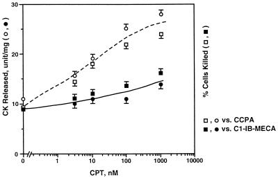
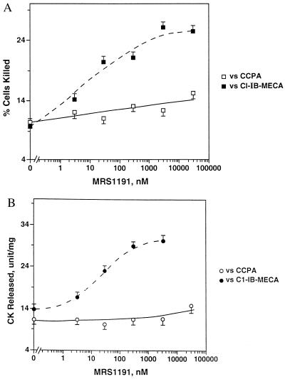
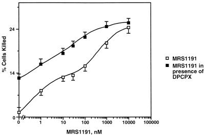
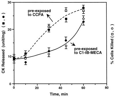
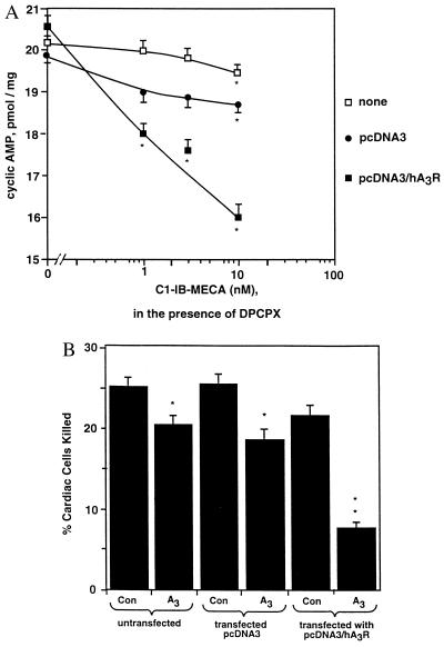
Adenosine released during cardiac ischemia exerts a potent, protective effect in the heart. A newly recognized adenosine receptor, the A3 subtype, is expressed on the cardiac ventricular cell, and its activation protects the ventricular heart cell against injury during a subsequent exposure to ischemia. A cultured chicken ventricular myocyte model was used to investigate the cardioprotective role of a novel adenosine A3 receptor. The protection mediated by prior activation of A3 receptors exhibits a significantly longer duration than that produced by activation of the adenosine A1 receptor. Prior exposure of the myocytes to brief ischemia also protected them against injury sustained during a subsequent exposure to prolonged ischemia. The adenosine A3 receptor-selective antagonist 3-ethyl 5-benzyl-2-methyl-6-phenyl-4-phenylethynyl-1, 4-(+/-)-dihydropyridine-3,5-dicarboxylate (MRS1191) caused a biphasic inhibition of the protective effect of the brief ischemia. The concomitant presence of the A1 receptor antagonist 8-cyclopentyl-1,3-dipropylxanthine (DPCPX) converted the MRS1191-induced dose inhibition curve to a monophasic one. The combined presence of both antagonists abolished the protective effect induced by the brief ischemia. Thus, activation of both A1 and A3 receptors is required to mediate the cardioprotective effect of the brief ischemia. Cardiac atrial cells lack native A3 receptors and exhibit a shorter duration of cardioprotection than do ventricular cells. Transfection of atrial cells with cDNA encoding the human adenosine A3 receptor causes a sustained A3 agonist-mediated cardioprotection. The study indicates that cardiac adenosine A3 receptor mediates a sustained cardioprotective function and represents a new cardiac therapeutic target.


Figures

Figure 1
Figure 1
Effect of 8-cyclopentyltheophylline on the CCPA- and Cl-IB-MECA-induced cardioprotective effect. Cultured ventricular cells were prepared and exposed to either CCPA (10 nM) or Cl-IB-MECA (10 nM) in the presence and absence of varying concentrations of the adenosine A1 receptor antagonist 8-cyclopentyltheophylline. Media were changed to that lacking agonist or antagonist. Cells were then exposed to room air at 37°C for 10 min before being exposed to simulated ischemia for 90 min. The percentage of cells killed and the amount of creatine kinase released were determined after the 90-min ischemic period. Data represented the means and standard errors of four experiments.
Figure 2
Figure 2
Effect of MRS1191 on the CCPA- and Cl-IB-MECA-induced cardioprotective effect. Cultured ventricular cells were prepared and exposed to either CCPA (10 nM) or Cl-IB-MECA (10 nM) in the presence and absence of varying concentrations of the adenosine A3 receptor antagonist MRS1191. Media were replaced with fresh media lacking the agonist or the antagonist. Cells were then exposed to normal O2 for 10 min before being exposed to 90 min of simulated ischemia. The percentage of myocytes killed (A) and the amount of creatine kinase released (B) were determined after the 90-min ischemic period. Data represented the means and standard errors of four experiments.
Figure 3
Figure 3
Adenosine A1 and A3 receptors mediate the cardioprotective effect of brief ischemia. Cultured ventricular cells were exposed to 5 min of simulated ischemia in the presence and the absence of various concentrations of MRS1191 or MRS1191 plus DPCPX (1 μM). Media were replaced by that lacking the receptor antagonists for 10 min before a 90-min ischemic exposure. The percentage of cells killed determined during the 90 min of ischemia represented the maximal level of ischemia-induced injury. Data were plotted as percentage of cells killed versus the concentration of MRS1191 in the presence and the absence of DPCPX. Data were means and standard errors of four experiments.
Figure 4
Figure 4
Differential cardioprotective duration mediated by the adenosine A1 and A3 receptors. Cultured ventricular cells were prepared and exposed to either CCPA (10 nM) or Cl-IB-MECA (10 nM) for 5 min. Media were replaced with fresh media lacking the agonist. Cells were then incubated under normal O2 for 10, 30, 45, or 60 min before being exposed to 90 min of simulated ischemia. The percentage of cells killed was determined after the 90-min ischemic period. Data represented the means and standard errors of five experiments. At the 30- and 45-min time points after the adenosine agonist exposure, the percentage of cells killed and the amount of creatine kinase released were significantly less in Cl-IB-MECA-treated than in CCPA-treated myocytes (t test, P < 0.01).
Figure 5
Figure 5
Acquisition of adenosine A3 receptor-mediated cyclic AMP and cardioprotective responses. Atrial cells were cultured from chicken embryos 14 days in ovo and transfected with pcDNA3 or with pcDNA3/hA3R (full-length cDNA encoding human adenosine A3 receptor subcloned in pcDNA3). (A) The ability of A3 receptor agonist Cl-IB-MECA to mediate inhibition of isoproterenol-stimulated cyclic AMP accumulation in untransfected atrial cells, in cells transfected with pcDNA3, or in cells transfected with pcDNA3/hA3R was determined. DPCPX (1 μM) was included to block the A1 receptor. Data were means and standard errors of triplicate determinations and were typical of five other experiments. ∗, The cyclic AMP level was significantly less than that obtained in cells not exposed to Cl-IB-MECA (control cells). At 10 nM of Cl-IB-MECA, the A3 receptor-mediated inhibition of the cyclic AMP level, expressed as percentage decrease from control cells, was significantly more in pcDNA3/hA3R-transfected cells than in pcDNA3-transfected cells or untransfected cells (one-way ANOVA and t test, P < 0.01). (B) The ability of Cl-IB-MECA to cause cardioprotection in untransfected atrial cells, in cells transfected with pcDNA3, or in cells transfected with pcDNA3/hA3R was determined. Cells were exposed to 10 nM of Cl-IB-MECA and 1 μM of DPCPX for 5 min. Media were replaced with fresh media lacking Cl-IB-MECA or DPCPX. Cells were then incubated under room air for 30 min before being exposed to 90 min of simulated ischemia. The percentage of myocytes killed and the amount of creatine kinase released (data not shown) were determined after the 90-min ischemic period. Data were the means and standard errors of seven experiments. In untransfected cells or in cells transfected with pcDNA3 or pcDNA3/hA3R, prior A3 agonist stimulation reduced the percentage of cells killed or the amount of CK released compared with cells not preexposed to A3 agonist (control cells) (∗, t test, P < 0.05). However, the A3 receptor-mediated reduction in the number of cells killed or the amount of CK released, expressed as percentage of decrease from those obtained in the control cells, was significantly more in pcDNA3/hA3R-transfected than in pcDNA3-transfected cells or untransfected cells (∗∗, one-way ANOVA and t test, P < 0.01).

Similar articles

Cited by

References

    1. Ely S W, Berne R M. Circulation. 1992;85:893–904. - PubMed
    1. Murry C E, Jennings R B, Reimer K A. Circulation. 1986;74:1124–1136. - PubMed
    1. Li G C, Vasquez J A, Gallagher K P, Lucchesi B R. Circulation. 1990;82:609–619. - PubMed
    1. Gross G J. Basic Res Cardiol. 1995;90:85–88. - PubMed
    1. Strickler J, Jacobson K A, Liang B T. J Clin Invest. 1996;98:1773–1779. - PMC - PubMed

Publication types

LinkOut - more resources