. 1998 Jun 9;95(12):7080-5.
doi: 10.1073/pnas.95.12.7080.

Aberrant intracellular localization of Varicella-Zoster virus regulatory proteins during latency

Affiliations

Aberrant intracellular localization of Varicella-Zoster virus regulatory proteins during latency

O Lungu et al. Proc Natl Acad Sci U S A. .

Abstract

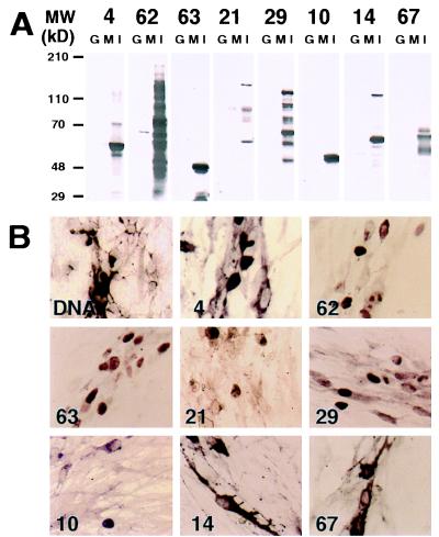
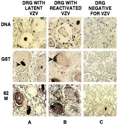
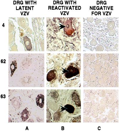
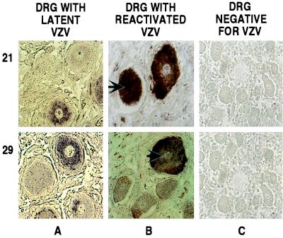
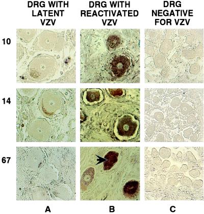
Varicella-Zoster virus (VZV) is a herpesvirus that becomes latent in sensory neurons after primary infection (chickenpox) and subsequently may reactivate to cause zoster. The mechanism by which this virus maintains latency, and the factors involved, are poorly understood. Here we demonstrate, by immunohistochemical analysis of ganglia obtained at autopsy from seropositive patients without clinical symptoms of VZV infection that viral regulatory proteins are present in latently infected neurons. These proteins, which localize to the nucleus of cells during lytic infection, predominantly are detected in the cytoplasm of latently infected neurons. The restriction of regulatory proteins from the nucleus of latently infected neurons might interrupt the cascade of virus gene expression that leads to a productive infection. Our findings raise the possibility that VZV has developed a novel mechanism for maintenance of latency that contrasts with the transcriptional repression that is associated with latency of herpes simplex virus, the prototypic alpha herpesvirus.


Figures

Figure 1
Figure 1
Specificity of the anti-VZV protein antibodies by Western blot analysis and in situ immunohistochemistry. (A) Purified bacterially expressed GST protein (0.5 μg, G), and VZV-infected (I) or uninfected (M) human embryonic lung fibroblast cell lysates were analyzed by SDS/PAGE on 7–12% gradient gels, and the proteins subsequently were transferred electrophoretically onto nitrocellulose membranes. The membranes were probed with the purified antibodies at a 1/1,000 dilution. The VZV ORFs whose products were analyzed are identified at the top. The molecular weights of prestained size markers (GIBCO/BRL, high molecular weight) are indicated on the left. (B) In situ hybridization and immunohistochemical detection of VZV DNA and proteins was performed in human embryonic lung fibroblasts infected with VZV strain Ellen. VZV DNA was detected by using a fluoresceine-labeled oligonucleotide probe and an AP-conjugated antifluorescein antibody. The products of ORFs 4, 62, 63, 21, 29, 10, 14, and 67 were detected by using purified anti-VZV proteins rabbit antibodies and AP-conjugated goat anti-rabbit Ig secondary antibody. The signal was visualized by developing with AP substrate. In situ DNA hybridization analysis is shown in the top left (DNA), and the ORFs whose products were analyzed are identified in the lower left corner of each panel.
Figure 2
Figure 2
In situ detection of VZV DNA and immunohistochemical analyses of VZV and GST proteins in human DRG. DRG harboring latent (A) or reactivated (B) virus and a control fetal DRG (C) were analyzed. VZV DNA and GST proteins were detected as described in the legend to Fig. 1. The product of VZV ORF62 was detected by using primary mouse mAbs, fluoresceine-labeled goat-anti-mouse secondary antibodies, and AP-conjugated goat-antifluoresceine tertiary antibody. The signal was visualized by developing with AP substrate. The specimens used are shown at the top, and the individual products analyzed are shown on the left. The large arrow indicates a neuron with a positive nucleus, and the small arrows point to coloration of lipofuschin.
Figure 3
Figure 3
Immunohistochemical detection of VZV IE proteins in human DRG. DRG harboring latent (A) or reactivated (B) virus and a control fetal DRG (C) were analyzed. The products of ORFs 4, 62, and 63 were detected as described in the legend to Fig. 1. The specimens used are shown at the top, and the individual genes whose products were analyzed are shown on the left. The arrows indicate neurons with positive nuclei. The staining seen near the plasma membrane of some neurons is from coloration of lipofuschin.
Figure 4
Figure 4
Immunohistochemical detection of VZV E proteins in human DRG. DRG harboring latent (A) or reactivated (B) virus and a control fetal DRG (C) were analyzed. The products of ORFs 21 and 29 were detected as described in the legend to Fig. 1. The specimens used are shown at the top, and the individual genes whose products were analyzed are shown on the left. The arrows indicate neurons with positive nuclei. The staining seen near the plasma membrane of some neurons is from coloration of lipofuschin.
Figure 5
Figure 5
Immunohistochemical detection of VZV late proteins in human DRG. DRG harboring latent (A) or reactivated (B) virus and a control fetal DRG (C) were analyzed. The products of ORFs 10, 14, and 67 were detected as described in the legend to Fig. 1. The specimens used are shown at the top, and the individual genes whose products were analyzed are shown on the left. The arrow indicates a neuron with a positive nucleus. The staining seen near the plasma membrane of some neurons is from coloration of lipofuschin.

References

    1. Garcia-Blanco M A, Cullen B R. Science. 1991;254:815–820. - PubMed
    1. Hope-Simpson R E. Proc R Soc Med. 1965;58:9–20. - PMC - PubMed
    1. Ragozzino M W, Melton L J, 3rd, Kurland L T, Chu C P, Perry H O. Medicine (Baltimore) 1982;51:310–316. - PubMed
    1. Straus S E. J Am Med Assoc. 1993;269:1836–1839. - PubMed
    1. Hyman R W, Ecker J R, Tenser R B. Lancet. 1983;2:814–816. - PubMed

Publication types