. 1998 Jun 9;95(12):7121-6.
doi: 10.1073/pnas.95.12.7121.

On the actions that one nerve cell can have on another: distinguishing "drivers" from "modulators"

Affiliations

On the actions that one nerve cell can have on another: distinguishing "drivers" from "modulators"

S M Sherman et al. Proc Natl Acad Sci U S A. .

Abstract

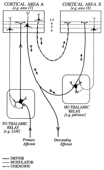
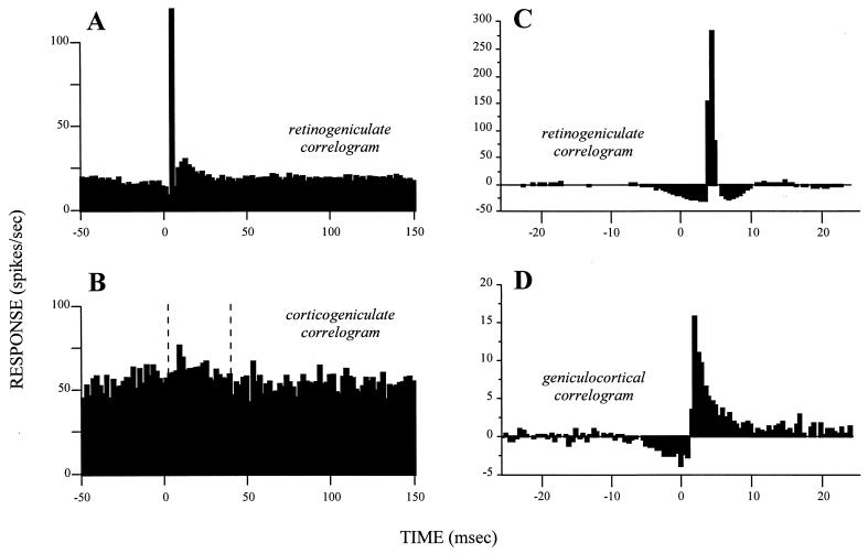
When one nerve cell acts on another, its postsynaptic effect can vary greatly. In sensory systems, inputs from "drivers" can be differentiated from those of "modulators." The driver can be identified as the transmitter of receptive field properties; the modulator can be identified as altering the probability of certain aspects of that transmission. Where receptive fields are not available, the distinction is more difficult and currently is undefined. We use the visual pathways, particularly the thalamic geniculate relay for which much relevant evidence is available, to explore ways in which drivers can be distinguished from modulators. The extent to which the distinction may apply first to other parts of the thalamus and then, possibly, to other parts of the brain is considered. We suggest the following distinctions: Cross-correlograms from driver inputs have sharper peaks than those from modulators; there are likely to be few drivers but many modulators for any one cell; and drivers are likely to act only through ionotropic receptors having a fast postsynaptic effect whereas modulators also are likely to activate metabotropic receptors having a slow and prolonged postsynaptic effect.


Figures

Figure 1
Figure 1
Schema to illustrate cortical and thalamic pathways. Two thalamic nuclei are shown: a first order (FO) relay on the left and a higher order (HO) relay on the right. A first order relay receives its driver inputs on proximal dendrites from subcortical sources via ascending pathways whereas a higher order relay receives its driver inputs from cells in cortical layer 5 (see ref. 40). The first order relay sends a driver input to layer 4 of cortical area A (thick line), and that same cortical area sends a modulator input (thin line with small terminals onto distal dendrites of the thalamic relay cell) from layer 6 back to the same first order thalamic nucleus. Cortical area A in turn sends a driver input from layer 5 to the higher order thalamic relay. This higher order relay sends its thalamocortical axons (shown as drivers, on the assumption that all thalamocortical inputs to layer 4 are drivers, although empirical data are lacking) to cortical area B and receives a modulator input back from layer 6 of cortical area B. Note that there are two paths by which cortical area A can influence area B. One is the transthalamic path, shown by small arrows and drawn as thick lines indicative of a driver pathway. The other is the direct corticocortical pathway (a “feed-forward” pathway, as defined by ref. 39), and this is shown by small arrows and as alternating thin and thick lines to indicate that we do not know whether this (or any other corticocortical pathway) represents a driver or a modulator input. As one specific example of such circuitry, we indicate the lateral geniculate nucleus (LGN) as a first order relay innervating area 17 and the pulvinar as a higher order relay receiving driver input from layer 5 of area 17 (41) and, in turn, innervating area 18.
Figure 2
Figure 2
Cross-correlograms displaying the difference between drivers and modulators. Each is based on simultaneous recordings in cats from two neurons, one presynaptic to the other. The cross-correlograms represent the firing of the postsynaptic cells relative to a spike at time zero for the presynaptic cell. (A) Retinogeniculate cross-correlogram based on spontaneous activity in both the retinal and geniculate neurons. Note the narrow peak rising out of a flat, low baseline that marks this as a driver connection. Redrawn from Fig. 3A of ref. , with permission of the publisher. (B) Corticogeniculate cross-correlogram based on spontaneous activity in both the layer 6 cell in area 17 and geniculate neuron. Glutamate was applied to the cortex to enhance the spontaneous firing of the afferent cell. Between the vertical, dashed lines, it is possible to discern a very gradual, prolonged, and small peak arising from a noisy, high baseline that marks this as a modulator connection. Redrawn from Fig. 2A of ref. , with permission of the publisher. (C and D) Cross-correlograms taken from the same laboratory by using identical techniques for easier comparison. Both are based on visually driven activity and involve a “shuffle correction” (43), and they are normalized against the firing level of the afferent, which is why some bins fall below zero. Both represent driver inputs and include another retinogeniculate pair (C) plus a geniculocortical pair (D). Note the difference in vertical scale, indicating that the retinal input accounts for more postsynaptic spikes in the geniculate cell (C) than does the geniculate input to the layer 4 cell of the striate cortex (D). Note also that the time represented by these cross-correlograms is much briefer than that for A and B. Nonetheless, both cross-correlograms have narrow peaks rising from a flat, low baseline, marking them as driver inputs. Data kindly provided by the authors for replotting. C is redrawn from data of Usrey et al.(15), and D is redrawn from Fig. 2 of Reid and Alonso (44).

Similar articles

Cited by

References

    1. Crick F, Koch C. Nature (London) 1998;391:245–250. - PubMed
    1. Feig S, Harting J K. J Comp Neurol. 1998;395:281–295. - PubMed
    1. Rockland K S. J Comp Neurol. 1997;390:515–536. - PubMed
    1. Sherman S M, Koch C. In: The Synaptic Organization of the Brain. Shepherd G M, editor. Vol. 4. London: Oxford Univ. Press; 1998. pp. 289–328.
    1. Sherman S M, Guillery R W. J Neurophysiol. 1996;76:1367–1395. - PubMed

Publication types

LinkOut - more resources