. 1998 Jun 9;95(12):7151-6.
doi: 10.1073/pnas.95.12.7151.

Bradykinin inhibits M current via phospholipase C and Ca2+ release from IP3-sensitive Ca2+ stores in rat sympathetic neurons

Affiliations

Bradykinin inhibits M current via phospholipase C and Ca2+ release from IP3-sensitive Ca2+ stores in rat sympathetic neurons

H Cruzblanca et al. Proc Natl Acad Sci U S A. .

Abstract

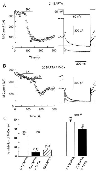
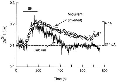
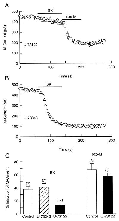
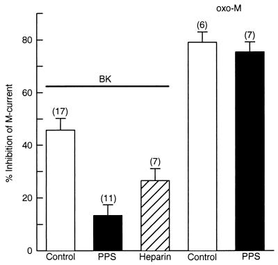
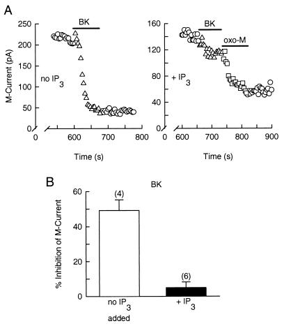
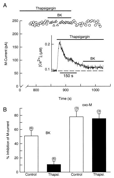
A variety of intracellular signaling pathways can modulate the properties of voltage-gated ion channels. Some of them are well characterized. However, the diffusible second messenger mediating suppression of M current via G protein-coupled receptors has not been identified. In superior cervical ganglion neurons, we find that the signaling pathways underlying M current inhibition by B2 bradykinin and M1 muscarinic receptors respond very differently to inhibitors. The bradykinin pathway was suppressed by the phospholipase C inhibitor U-73122, by blocking the IP3 receptor with pentosan polysulfate or heparin, and by buffering intracellular calcium, and it was occluded by allowing IP3 to diffuse into the cytoplasm via a patch pipette. By contrast, the muscarinic pathway was not disrupted by any of these treatments. The addition of bradykinin was accompanied by a [Ca2+]i rise with a similar onset and time to peak as the inhibition of M current. The M current inhibition and the rise of [Ca2+]i were blocked by depletion of Ca2+ internal stores by thapsigargin. We conclude that bradykinin receptors inhibit M current of sympathetic neurons by activating phospholipase C and releasing Ca2+ from IP3-sensitive Ca2+ stores, whereas muscarinic receptors do not use the phospholipase C pathway to inhibit M current channels.


Figures

Figure 1
Figure 1
Intracellular BAPTA blocks modulation of M current by BK. Symbols represent the amplitude of M current deactivation measured every 4 s. BK and oxo-M were applied as indicated by the horizontal bars. (A) BK elicits a strong inhibition of M current (triangles) in a neuron dialyzed with 0.1 mM BAPTA. Voltage protocol and M current recording for the control (a) and during BK exposure (b) are shown on the right. Dashed line is the zero current level. (B) BK (triangles), but not oxo-M (squares), fails to inhibit M current in a neuron dialyzed with a 20 mM BAPTA/10 mM Ca2+ pipette solution. M current records from the control condition (a), with BK (b) and oxo-M (c) shown on the right. (C) Bar plot of the mean suppression (±SEM) of M current by BK (50 nM) and oxo-M (10 μM) with different internal solutions. The number of cells tested for each condition is in parentheses. M current records in the high 20 mM BAPTA pipette solutions were taken 9 min after breakthrough of the patch membrane.
Figure 2
Figure 2
BK raises [Ca2+]i to inhibit M current. Simultaneous recordings of [Ca2+]i and M current were done in a neuron loaded with indo-1 dye through the patch pipette (0.1 mM BAPTA/0.1 mM indo-1). In this neuron, BK raised [Ca2+]i (continuous trace) from 105 nM to 218 nM. The dashed line is the basal [Ca2+]i level. For comparison, the M current data (circles) were inverted (vertical bar) and aligned to match the amplitude of the [Ca2+]i signal.
Figure 3
Figure 3
Suppression of M current by BK is attenuated by the PLC inhibitor U-73122. Cells were incubated for 30 min with U-73122 (1 μM) or U-73343 (1 μM). BK (100 nM) and oxo-M (5 μM) were applied as indicated by the horizontal bars. (A) U-73122 prevents M current suppression by BK (triangles) but not by oxo-M (squares). (B) The inactive analog U-73343 does not block modulation of M current by BK. (C) Mean (±SEM) inhibition of M current by BK or by oxo-M in control cells (open bars) and in neurons treated with U-73343 (dashed bar) or U-73122 (solid bars).
Figure 4
Figure 4
Antagonists of the IP3 receptor selectively disrupt the BK pathway. Neurons were dialyzed with PPS or heparin for 14 min before M current recording. Mean (±SEM) inhibition of M current by BK (100 nM) and oxo-M (10 μM) in control cells (open bars) and in neurons dialyzed with PPS (solid bars) or heparin (dashed bar). PPS (100 μg/ml) disrupts M current inhibition by BK but not by oxo-M. Heparin (200 μg/ml) was less effective than PPS to block the BK-activated pathway.
Figure 5
Figure 5
IP3 occludes the inhibitory action of BK on M current. M current records were taken 10 min after breakthrough of the patch membrane. (A Left) In a control cell, BK suppresses M current. (A Right) In a neuron dialyzed with IP3 (100 μM), BK does not inhibit M current well (triangles), whereas oxo-M does (squares). (B) Mean inhibition (±SEM) of M current by BK in control (open bar) and IP3-loaded cells (solid bar).
Figure 6
Figure 6
Thapsigargin prevents the BK-evoked increase of [Ca2+]i and suppression of M current. (A) In a cell treated with thapsigargin (5 μM) for 13 min, BK does not inhibit M current. (Inset) Thapsigargin transiently raises [Ca2+]i from 92 to 210 nM and blocks a further rise of [Ca2+]i by BK (data from a different cell). (B) Mean (±SEM) M current inhibition by BK (100 nM) or oxo-M (10 μM) in control cells (open bars) and in neurons treated with thapsigargin (Thapsi., solid bars).

References

    1. Hille B. Trends Neurosci. 1994;17:531–536. - PubMed
    1. Bernheim L, Beech D J, Hille B. Neuron. 1991;6:859–867. - PubMed
    1. Shapiro M S, Wollmuth L P, Hille B. Neuron. 1994;12:1319–1329. - PubMed
    1. Lewis D L, Ikeda S R. Neuroendocrinology. 1997;66:235–245. - PubMed
    1. Lee S B, Rhee S G. Curr Opin Cell Biol. 1995;7:183–189. - PubMed

Publication types

MeSH terms

LinkOut - more resources