. 1998 Jun 9;95(12):7174-8.
doi: 10.1073/pnas.95.12.7174.

A release mechanism for stored ATP in ocular ciliary epithelial cells

Affiliations

A release mechanism for stored ATP in ocular ciliary epithelial cells

C H Mitchell et al. Proc Natl Acad Sci U S A. .

Abstract

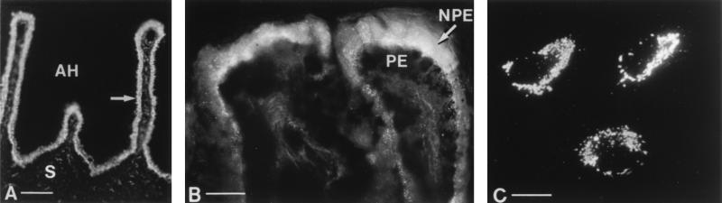
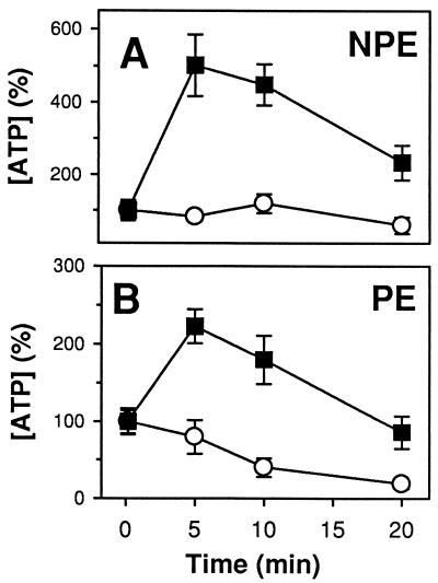
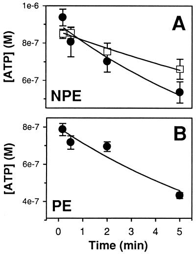
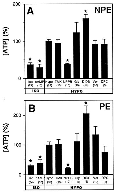
Purines can modify ciliary epithelial secretion of aqueous humor into the eye. The source of the purinergic agonists acting in the ciliary epithelium, as in many epithelial tissues, is unknown. We found that the fluorescent ATP marker quinacrine stained rabbit and bovine ciliary epithelia but not the nerve fibers in the ciliary bodies. Cultured bovine pigmented and nonpigmented ciliary epithelial cells also stained intensely when incubated with quinacrine. Hypotonic stimulation of cultured epithelial cells increased the extracellular ATP concentration by 3-fold; this measurement underestimates actual release as the cells also displayed ecto-ATPase activity. The hypotonically triggered increase in ATP was inhibited by the Cl--channel blocker 5-nitro-2-(3-phenylpropylamino)benzoic acid (NPPB) in both cell types. In contrast, the P-glycoprotein inhibitors tamoxifen and verapamil and the cystic fibrosis transmembrane conductance regulator (CFTR) blockers glybenclamide and diphenylamine-2-carboxylate did not affect ATP release from either cell type. This pharmacological profile suggests that ATP release is not restricted to P-glycoprotein or the cystic fibrosis transmembrane conductance regulator, but can proceed through a route sensitive to NPPB. ATP release also was triggered by ionomycin through a different NPPB-insensitive mechanism, inhibitable by the calcium/calmodulin-activated kinase II inhibitor KN-62. Thus, both layers of the ciliary epithelium store and release ATP, and purines likely modulate aqueous humor flow by paracrine and/or autocrine mechanisms within the two cell layers of this epithelium.


Figures

Figure 1
Figure 1
Quinacrine staining in ciliary epithelial cells. (A) PE and NPE cell layers fluoresced brightly (arrow) in tissue sections of the ciliary body of an albino rabbit injected i.p. with quinacrine. The PE layer lies closest to the stroma (S) whereas the adjacent NPE layer faces the aqueous humor (AH); both layers stain intensely. Both layers of the ciliary epithelium from rabbits injected i.v. with quinacrine, or incubated in quinacrine also stained intensely (not shown). (B) The NPE cells of bovine tissue incubated with quinacrine showed particulate staining that was brightest on the apical membrane of the cells, facing the PE cells. PE cells did not clearly display fluorescent granules, but the densely packed melanin granules could have obscured the dye. (C) Particulate staining was observed in melanin-free cultured PE cells incubated in quinacrine. Cultured NPE cells (not shown) showed a similar pattern and intensity of staining. (Magnification bars: A, 100 μm; B and C, 25 μm.)
Figure 2
Figure 2
Hypotonically triggered release of ATP from cultured bovine NPE (A) and PE (B) cells. Extracellular samples obtained from cells bathed in hypotonic solution (▪) showed an increase in ATP concentration whereas samples from cells bathed in isotonic solution (○) did not. ATP was normalized to the 10-sec isosmotic measurement for each set of experiments. Separate dishes were used for each time measurement (n = 5–10).
Figure 3
Figure 3
Degradation of externally added ATP by ciliary epithelial cells. ATP (1.2 μM) in isotonic solution was added to dishes of NPE (A) or PE (B) cells at t = 0, and a sample of extracellular solution was removed at the time indicated. Separate dishes were used for each time point. ATP was degraded at a lower rate (P < 0.05, n = 4–5) in isotonic containing only 1 μM Ca2+ and Mg2+ (A, □, τ = 19 ± 1 min) rather than 1.3 mM Ca2+ and 0.5 mM Mg2+ (A, •, τ = 8 ± 1 min; B, τ = 9 ± 2 min).
Figure 4
Figure 4
Manipulation of ATP release from cultured NPE (A) and PE (B) cells. ATP release in isotonic solution (Iso) was not triggered by a cAMP-stimulating cocktail. Hypotonic solution (Hypo) led to an increase in ATP that was blocked by 100 μM NPPB, but unaffected by 10 μM tamoxifen (TMX), 100 μM glybenclamide (Gly), 100 μM verapamil (Ver), or 500 μM DPC. DIDS (100 μM) increased ATP levels. (∗ = different from hypotonic levels, P < 0.001). The vehicles had no significant effect. ATP concentration was normalized to the mean hypotonic level for each day’s experiments.

Similar articles

Cited by

References

    1. Krupin T, Civan M M. In: The Glaucomas. 2nd Ed. Ritch R, Shields M B, Krupin T, editors. St. Louis: Mosby; 1995. pp. 251–280.
    1. Bowler J M, Peart D, Purves R D, Carré D A, Macknight A D C, Civan M M. Exp Eye Res. 1996;62:131–139. - PubMed
    1. Civan M M. News Physiol Sci. 1997;12:158–162.
    1. Wax M, Sanghavi D M, Lee C H, Kapadia M. Exp Eye Res. 1993;57:89–95. - PubMed
    1. Carré D A, Mitchell C H, Peterson-Yantorno K, Coca-Prados M, Civan M M. Am J Physiol. 1997;273:C1354–C1361. - PubMed

Publication types

MeSH terms

Substances

LinkOut - more resources