. 1998 Jun;36(6):1666-73.
doi: 10.1128/JCM.36.6.1666-1673.1998.

Cloning and expression of the 44-kilodalton major outer membrane protein gene of the human granulocytic ehrlichiosis agent and application of the recombinant protein to serodiagnosis

Affiliations

Cloning and expression of the 44-kilodalton major outer membrane protein gene of the human granulocytic ehrlichiosis agent and application of the recombinant protein to serodiagnosis

N Zhi et al. J Clin Microbiol. 1998 Jun.

Erratum in

Abstract

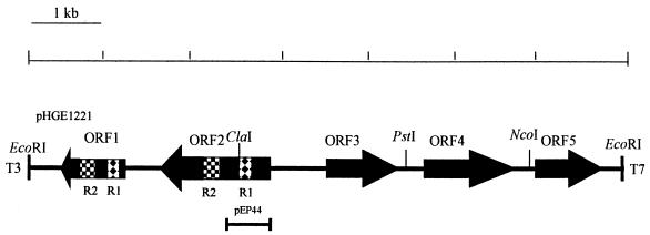
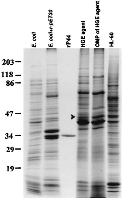
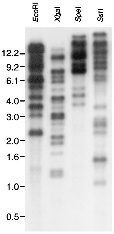
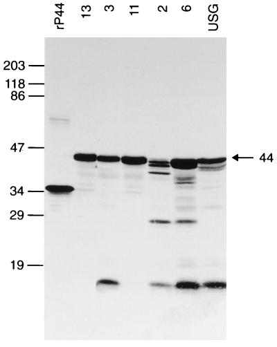
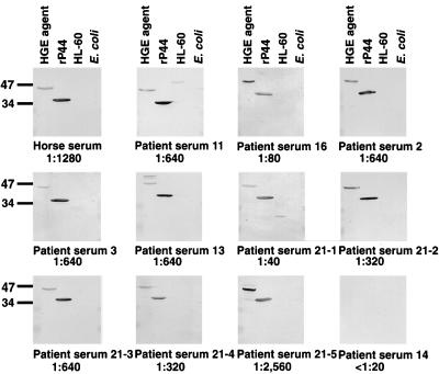
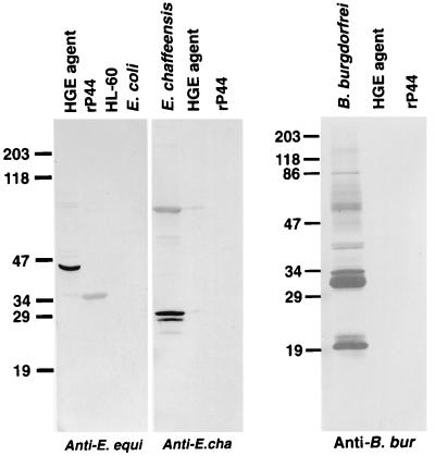
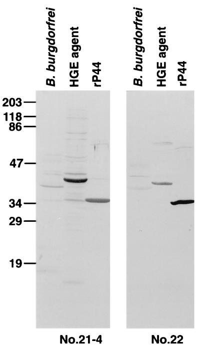
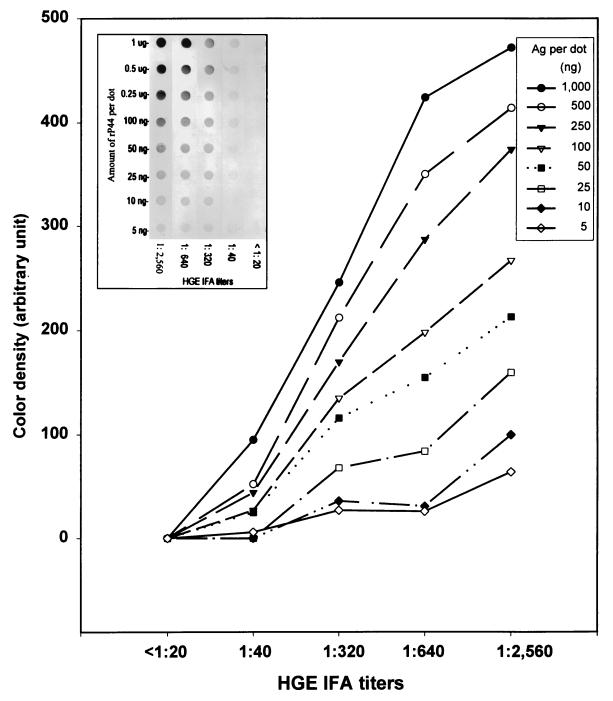
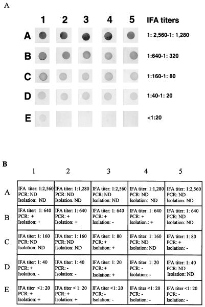
A 44-kDa major outer membrane protein of the human granulocytic ehrlichiosis (HGE) agent is an immunodominant antigen in human infection. A gene encoding this protein was cloned and sequenced. Southern blot results revealed the existence of multigenes homologous to the P44 gene in the genome of the HGE agent. The recombinant 44-kDa protein (rP44) was expressed by using expression vector pET30a. The reactivity of the affinity-purified rP44 was evaluated by Western immunoblot analysis and dot blot immunoassay. Western immunoblot analysis showed that mouse anti-rP44 serum reacted with 44- to 42-kDa proteins in six different HGE agent strains tested except strain 2, in which three proteins of 42, 40, and 38 kDa were recognized. Eleven HGE patient serum samples, a horse anti-HGE serum, and a horse anti-Ehrlichia equi serum recognized the rP44 protein. This suggests that rP44 is an HGE-E. equi group-specific antigen. Neither human anti-Ehrlichia chaffeensis serum nor rabbit anti-Borrelia burgdorferi serum reacted with rP44. Sera from two patients coinfected with the HGE agent and B. burgdorferi reacted positively with rP44 and the HGE agent. Sera from 20 HGE patients with indirect fluorescent-antibody (IFA) titers ranging from 1:20 to 1:2,560 gave distinct positive reactions in a dot immunoblot assay. There was a positive correlation between the color densities of the dot reactions and the IFA titers when greater than 50 ng of recombinant antigen per dot was used. The use of the affinity-purified rP44 protein as antigen would provide a more specific, consistent, and simpler serodiagnosis for HGE than the use of whole infected cells or purified HGE agents.


Figures

FIG. 1
FIG. 1
Restriction map of a 6.9-kb genomic DNA fragment including the P44 gene of the HGE agent. The closed boxes with arrows indicate the four ORFs that are identified in this fragment. The arrows indicate the orientations of these ORFs. Patterned boxes (R1 and R2) show two identical regions in ORF1 which encoded 59 and 65 amino acids, respectively. The solid bar at the bottom indicates the region which was cloned into the pET30a expression vector.
FIG. 2
FIG. 2
SDS-PAGE patterns of the antigens used in this study. The proteins (10 μg) were separated in an SDS–10% polyacrylamide gel and stained with Coomassie blue. The arrowhead indicates the native 44-kDa protein in the whole-cell organisms and outer membrane protein fraction (OMP) of the HGE agent. The numbers on the left indicate the molecular masses in kilodaltons based on the broad-range prestained standards (Bio-Rad).
FIG. 3
FIG. 3
Genomic Southern blot analysis of the HGE agent isolate 13 with a 32P-labeled 1.2-kb P44 gene probe. The numbers on the left indicate molecular sizes in kilobases.
FIG. 4
FIG. 4
Western immunoblot analysis of mouse anti-rP44 serum using six different HGE agent isolates and the rP44 antigen. Samples subjected to SDS-PAGE consisted of 10 μg of purified whole-cell preparation of the HGE agent isolates 13, 3, 11, 2, 6, and USG and affinity-purified rP44. These proteins were transferred to a nitrocellulose sheet and incubated with a 1:1,000 dilution of antisera. The numbers on the left indicate molecular masses in kilodaltons based on the broad-range prestained standards (Bio-Rad).
FIG. 5
FIG. 5
Western immunoblot analysis of anti-HGE sera using purified HGE agent isolate 13 and rP44 antigen. The sera used in this study included a horse anti-HGE agent serum, five convalescent-phase serum samples from patients 2, 3, 11, 13, and 16 (23), five serum samples collected at different times of illness over a 2-year period from patient 21, who was suspected of having a persistent infection or reinfection, and negative control serum (IFA titer, <1:20). Samples subjected to SDS-PAGE consisted of 10 μg of purified whole-cell preparation of HGE agent strain 13 (23), affinity-purified rP44, HL-60 cells, and E. coli BL21(DE3)/pLysS. These proteins were transferred to a nitrocellulose sheet and incubated with a 1:1,000 dilution of antisera. The number at the bottom of each panel represent the IFA test titer of the serum sample. The numbers on the left indicate molecular masses in kilodaltons based on the broad-range prestained standards (Bio-Rad).
FIG. 6
FIG. 6
Western immunoblot analysis of rP44 antigen of the HGE agent with sera against E. equi, E. chaffeensis, and B. burgdorferi. The sera used in this study included horse anti-E. equi serum, human anti-E. chaffeensis serum, and rabbit anti-B. burgdorferi serum. Antigens subjected to SDS-PAGE were 10 μg of purified whole-cell preparation of HGE agent isolate 13, E. chaffeensis, or B. burgdorferi, affinity-purified rP44, uninfected HL-60 cells, and E. coli BL21(DE3)/pLysS. These proteins were transferred to a nitrocellulose sheet and incubated with a 1:1,000 dilution of antisera. The numbers on the left indicate molecular masses in kilodaltons based on the broad-range prestained standards (Bio-Rad).
FIG. 7
FIG. 7
Western immunoblot analysis of rP44 antigen of the HGE agent with serum samples from patients with HGE-Lyme borreliosis coinfection. Serum samples 21-5 and 22 used in this study were collected from patients who were diagnosed as coinfected with HGE agent and B. burgdorferi. Antigens subjected to SDS-PAGE were 10 μg of purified whole-cell preparation of HGE agent isolate 13, B. burgdorferi, and affinity-purified rP44. These proteins were transferred to a nitrocellulose sheet and incubated with a 1:1,000 dilution of antisera. Numbers on the left indicate the molecular masses in kilodaltons based on the broad-range prestained standards (Bio-Rad).
FIG. 8
FIG. 8
Optical density analysis of the reaction of the HGE agent rP44 antigen (Ag) with patient sera having different IFA testing titers was performed by using ImageQuaNT computer program. Color development of various amounts of affinity-purified rP44 of the HGE agent was done after reaction with patient sera having different HGE agent IFA titers and with negative control sera (insert). The sera were diluted at 1:1,000.
FIG. 9
FIG. 9
(A) Dot blot immunoassay of HGE patient sera with different IFA testing titers by using 0.5 μg of affinity-purified rP44 antigen. The sera were diluted at 1:1,000. (B) Other diagnostic data for the blood serum samples used in this assay are shown. Symbols: −, negative; +, positive. ND, not done.

Similar articles

Cited by

References

    1. Aguero-Rosenfeld M E, Horowitz H W, Wormser G P, Mckenna D F, Nowakowski J, Munoz J, Dumler J S. Human granulocytic ehrlichiosis: a case series from a medical center in New York State. Ann Intern Med. 1996;125:904–908. - PubMed
    1. Alleman A R, Palmer G H, McGuire T C, McElwain T F, Perryman L E, Barbet A F. Anaplasma marginale major surface protein 3 is encoded by a polymorphic, multigene family. Infect Immun. 1997;65:156–163. - PMC - PubMed
    1. Altschul S F, Gish W, Miller W, Myers E W, Lipman D J. Basic local alignment search tool. J Mol Biol. 1990;215:403–410. - PubMed
    1. Asanovich K M, Bakken J S, Madigan J E, Aguero-Rosenfeld M, Wormser G P, Dumler J S. Antigenic diversity of granulocytic Ehrlichia isolates from humans in Wisconsin and New York and a horse in California. J Infect Dis. 1997;176:1029–1034. - PubMed
    1. Bakken J S, Krueth J, Tilden R L, Dumler J S, Kristansen B E. Serological evidence of human granulocytic ehrlichiosis in Norway. Eur J Clin Microbiol Infect Dis. 1997;15:829–832. - PubMed

Publication types

MeSH terms

Associated data

LinkOut - more resources