

Invasive infection with Fusarium chlamydosporum in a patient with aplastic anemia
- PMID: 9620419
- PMCID: PMC104919
- DOI: 10.1128/JCM.36.6.1772-1776.1998
Invasive infection with Fusarium chlamydosporum in a patient with aplastic anemia
Abstract
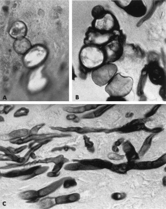
We report the first case of invasive disease caused by Fusarium chlamydosporum. The patient had aplastic anemia with prolonged neutropenia and was treated with immunosuppressive therapy. While she was receiving empirical amphotericin B, a dark crusted lesion developed on her nasal turbinate. Histologic analysis revealed invasive hyaline hyphae and some darkly pigmented structures that resembled conidia of dematiaceous molds. Only after the mold was grown in culture were characteristic colonial morphology, phialides, conidia, and chlamydospores evident, thus permitting the identification of F. chlamydosporum. This case illustrates the ever-increasing spectrum of pathogenic Fusarium spp. in immunocompromised patients and emphasizes the potential pitfalls in histologic diagnosis, which may have important treatment implications.
Figures

Similar articles
-
Fusarium infections in patients with severe aplastic anemia: review and implications for management.Haematologica. 1999 Feb;84(2):114-8. Haematologica. 1999. PMID: 10091408 Review.
-
Evaluation of Fusarium solani hyphae and conidia susceptibility to amphotericin B and itraconazole: study of a clinical case.Mycopathologia. 2005 Nov;160(4):291-6. doi: 10.1007/s11046-005-0106-2. Mycopathologia. 2005. PMID: 16244897
-
Disseminated infection caused by Fusarium solani in a patient with aplastic anemia.N Y State J Med. 1990 Dec;90(12):609-10. N Y State J Med. 1990. PMID: 2277679 No abstract available.
-
Invasive Exserohilum sinusitis in a patient with aplastic anemia.Pediatr Infect Dis J. 2005 Oct;24(10):939-41. doi: 10.1097/01.inf.0000180988.96528.5c. Pediatr Infect Dis J. 2005. PMID: 16220103
-
Invasive fungal sinusitis in patients undergoing bone marrow transplantation.Bone Marrow Transplant. 1993 Sep;12(3):203-8. Bone Marrow Transplant. 1993. PMID: 8241977 Review.
Cited by
-
Fusarium infections in immunocompromised patients.Clin Microbiol Rev. 2007 Oct;20(4):695-704. doi: 10.1128/CMR.00014-07. Clin Microbiol Rev. 2007. PMID: 17934079 Free PMC article. Review.
-
Internet-accessible DNA sequence database for identifying fusaria from human and animal infections.J Clin Microbiol. 2010 Oct;48(10):3708-18. doi: 10.1128/JCM.00989-10. Epub 2010 Aug 4. J Clin Microbiol. 2010. PMID: 20686083 Free PMC article.
-
Disseminated fusariosis with ecthyma gangrenosum-like lesions in a refractory acute myeloid leukemia patient.Curr Med Mycol. 2019 Mar;5(1):27-31. doi: 10.18502/cmm.5.1.534. Curr Med Mycol. 2019. PMID: 31049455 Free PMC article.
-
Hybrid de novo genome assembly data and comparative genomics of Fusarium chlamydosporum isolated from infected blackberry fields.Data Brief. 2025 Jul 9;61:111854. doi: 10.1016/j.dib.2025.111854. eCollection 2025 Aug. Data Brief. 2025. PMID: 40718146 Free PMC article.
-
Fusarium verticillioides abscess of the nasal septum in an immunosuppressed child: case report and identification of the morphologically atypical fungal strain.J Clin Microbiol. 2005 Apr;43(4):1998-2001. doi: 10.1128/JCM.43.4.1998-2001.2004. J Clin Microbiol. 2005. PMID: 15815043 Free PMC article.
References
-
- Anaissie E, Bodey G P, Kantarjian H, Ro J, Vartivarian S E, Hopfer R, Hoy J, Rolston K. New spectrum of fungal infections in patients with cancer. Rev Infect Dis. 1989;11:369–378. - PubMed
-
- Anaissie E, Kantarjian H, Jones P, Barlogie B, Luna M, Lopez-Berestein G, Bodey G P. Fusarium: a newly recognized fungal pathogen in immunosuppressed patients. Cancer. 1986;57:2141–2145. - PubMed
-
- Anaissie E, Kantarjian H, Ro J, Hopfer R, Rolston K, Fainstein V, Bodey G. The emerging role of Fusarium infections in patients with cancer. Medicine. 1988;67:77–83. - PubMed
-
- Anaissie E J, Bodey G P, Rinaldi M G. Emerging fungal infections. Eur J Clin Microbiol Infect Dis. 1989;8:323–330. - PubMed
-
- Boutati E I, Anaissie E J. Fusarium, a significant emerging pathogen in patients with hematologic malignancy: ten years’ experience at a cancer center and implications for management. Blood. 1997;90:999–1008. - PubMed
Publication types
MeSH terms
Substances
LinkOut - more resources
Full Text Sources
Medical
Miscellaneous
