. 1998 Jun 1;12(11):1593-8.
doi: 10.1101/gad.12.11.1593.

RNA-binding protein conserved in both microtubule- and microfilament-based RNA localization

Affiliations

RNA-binding protein conserved in both microtubule- and microfilament-based RNA localization

L Havin et al. Genes Dev. .

Abstract

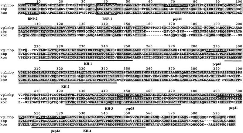
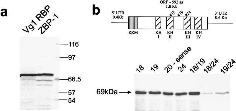
Vg1 mRNA translocation to the vegetal cortex of Xenopus oocytes requires intact microtubules, and a 3' UTR cis-acting element (termed VLE), which also mediates sequence-specific binding of several proteins. One protein, the 69-kD Vg1 RBP, associates Vg1 RNA to microtubules in vitro. Here we show that Vg1 RBP-binding sites correlate with vegetal localization. Purification and cloning of Vg1 RBP revealed five RNA-binding motifs: four KH and one RRM domains. Surprisingly, Vg1 RBP is highly homologous to the zipcode binding protein implicated in the microfilament-mediated localization of beta actin mRNA in fibroblasts. These data support Vg1 RBP's direct role in vegetal localization and suggest the existence of a general, evolutionarily conserved mechanism for mRNA targeting.


Figures

Figure 1
Figure 1
Cis-acting binding elements in the VLE and their role in localization. (a) The ability of different fragments of the VLE, as well as of the LS, substituted VLE fragments, to bind Vg1 RBP is schematically represented. The coordinates of the fragments and the substitution used for each binding assay (with the first nucleotide of the VLE numbered 1 and the substitution indicated as Ins) are listed at the left. The two regions identified as being important for Vg1 RBP binding are indicated by the boxes at the top. Domains protected by oocyte proteins from RNase degradation, mapped and labeled by Mowry (1996) as C, A/B, and D, respectively, are shown as lines beneath the full-length VLE. The degree of binding is indicated in the inset. (b) A representative UV cross-linking experiment (summarized in a) is shown for wild-type VLE, 1-366/ins1–20 (mut 1), 1-366/ins256–275 (mut 2), and 1–366/ins1-20&256–275 (mut 1&2) RNAs. (c) The nucleotide sequences of the first and second binding elements in the VLE are compared with a sequence from the 3′ UTR of Xcat-2 RNA from the region mapped as containing a late pathway localization element (Zhou and King 1996b). The hexanucleotide UUUCUA present in all these sequences is indicated in boldface type.
Figure 1
Figure 1
Cis-acting binding elements in the VLE and their role in localization. (a) The ability of different fragments of the VLE, as well as of the LS, substituted VLE fragments, to bind Vg1 RBP is schematically represented. The coordinates of the fragments and the substitution used for each binding assay (with the first nucleotide of the VLE numbered 1 and the substitution indicated as Ins) are listed at the left. The two regions identified as being important for Vg1 RBP binding are indicated by the boxes at the top. Domains protected by oocyte proteins from RNase degradation, mapped and labeled by Mowry (1996) as C, A/B, and D, respectively, are shown as lines beneath the full-length VLE. The degree of binding is indicated in the inset. (b) A representative UV cross-linking experiment (summarized in a) is shown for wild-type VLE, 1-366/ins1–20 (mut 1), 1-366/ins256–275 (mut 2), and 1–366/ins1-20&256–275 (mut 1&2) RNAs. (c) The nucleotide sequences of the first and second binding elements in the VLE are compared with a sequence from the 3′ UTR of Xcat-2 RNA from the region mapped as containing a late pathway localization element (Zhou and King 1996b). The hexanucleotide UUUCUA present in all these sequences is indicated in boldface type.
Figure 2
Figure 2
Localization of wild-type and mutant VLEs in oocytes. (a) The distribution of wild-type VLE RNA or double substituted VLE mut 1&2 RNA (1–366/ins1–20&256–275, see Fig. 1a for map) following injection into late stage III oocytes. mut 1&2 RNA appears to be fairly uniformly distributed throughout the oocyte; wild-type VLE shows strong cortical localization to the vegetal hemisphere alone. (b) Confocal micrographs of late stage III oocytes coinjected with wild-type VLE RNA (green channel) and either mut 1 RNA (1–366/ins1–20, see Fig. 1a) or mut 2 RNA (1–366/ins256–275) (red channel). Although the majority of both the mutant RNAs is not localized, some colocalization with the wild-type VLE RNA (yellow, third column) is observable.
Figure 3
Figure 3
Biochemical purification Vg1 RBP. (a) Fractions of the heparin–Sepharose column were assayed for Vg1 RBP cross-linking activity. KCl concentrations (as determined by conductivity) of selected fractions are indicated. (b) Equivalent fractions of the RNA affinity column were assayed for Vg1 RBP cross-linking activity (autoradiograph, top) and presence of protein (silver-stained gel, bottom). (Arrows) The location of Vg1 RBP.
Figure 4
Figure 4
Vg1 RBP protein sequence and its homology to ZBP-1 and KOC. The protein sequence for Vg1 RBP is shown with the four KH (underlined) and one RRM (boxed) domains indicated. The five peptides that were obtained from the microsequencing of the purified Vg1 RBP are underlined and labeled. ZBP-1 and KOC sequences are also shown, aligned to Vg1 RBP, and identical amino acids are shaded. Percent identities/homologies between the proteins are Vg1 RBP/ZBP-1, 78%/84%; Vg1 RBP/KOC, 83%/87%; ZBP-1/KOC, 76%/80%.
Figure 5
Figure 5
The cloned gene encodes Vg1 RBP. (a) In vitro translation of Vg1 RBP and ZBP-1 RNA. Both Vg1 RBP and ZBP-1 translation products migrate close to 69 kD, according to the molecular mass markers (right). (b) UV cross-linking of S100 extracts from oocytes injected with the indicated oligonucleotides was performed. Using a PhosphorImager to quantify the band intensities, we observed a 70% reduction in cross-linking activity (as compared to the control sense oligonucleotide 20) when oligonucleotides 18 and 24 are injected simultaneously, and a 50% reduction when 19 and 24 are injected together. The third pair of oligonucleotides (18 and 19) had no effect on Vg1 RBP-binding activity. The schematic drawing indicates the position of the oligonucleotides relative to the RNA-binding domains.

References

    1. Bassell GJ, Zhang H, Byrd AL, Femino AM, Singer RH, Taneja KL, Lifshitz LM, Herman IM, Kosik KS. Sorting of β-actin mRNA and protein to neurites and growth cones in culture. J Neurosci. 1998;18:251–265. - PMC - PubMed
    1. Deshler JO, Highett MI, Schnapp BJ. Localization of Xenopus Vg1 mRNA by Vera protein and the endoplasmic reticulum. Science. 1997;276:1128–1131. - PubMed
    1. Elisha Z, Havin L, Ringel I, Yisraeli JK. Vg1 RNA binding protein mediates the association of Vg1 RNA with microtubules in Xenopus oocytes. EMBO J. 1995;14:5109–5114. - PMC - PubMed
    1. Erdelyi M, Michon AM, Guichet A, Glotzer JB, Ephrussi A. Requirement for Drosophila cytoplasmic tropomyosin in oskar mRNA localization. Nature. 1995;377:524–527. - PubMed
    1. Forristall C, Pondel M, Chen L, King ML. Patterns of localization and cytoskeletal association of two vegetally localized RNAs, Vg1 and Xcat-2. Development. 1995;121:201–208. - PubMed

Publication types

MeSH terms

LinkOut - more resources