. 1998 Jun 1;12(11):1610-20.
doi: 10.1101/gad.12.11.1610.

Targeted disruption of SHIP leads to hemopoietic perturbations, lung pathology, and a shortened life span

Affiliations

Targeted disruption of SHIP leads to hemopoietic perturbations, lung pathology, and a shortened life span

C D Helgason et al. Genes Dev. .

Abstract

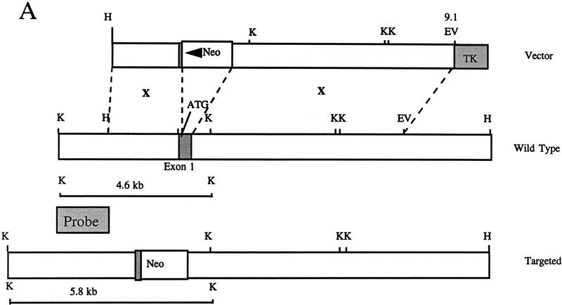
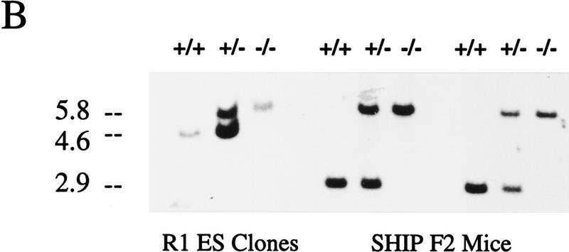
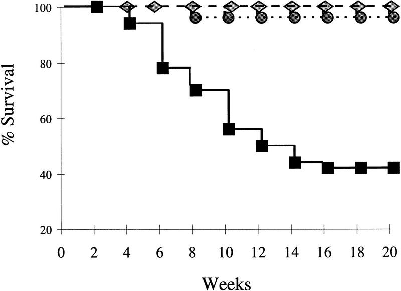
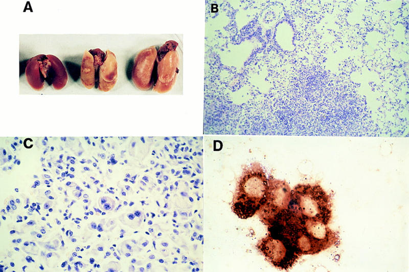
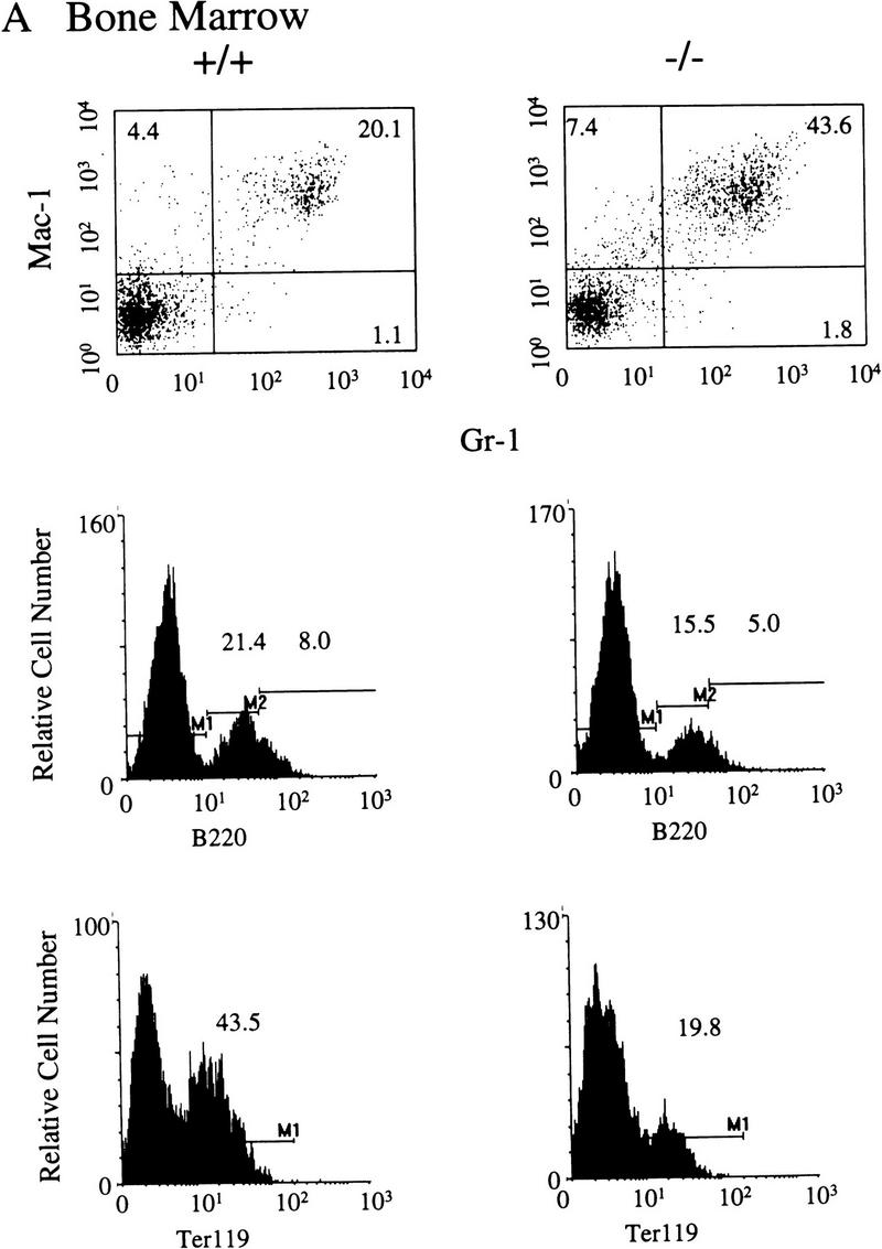
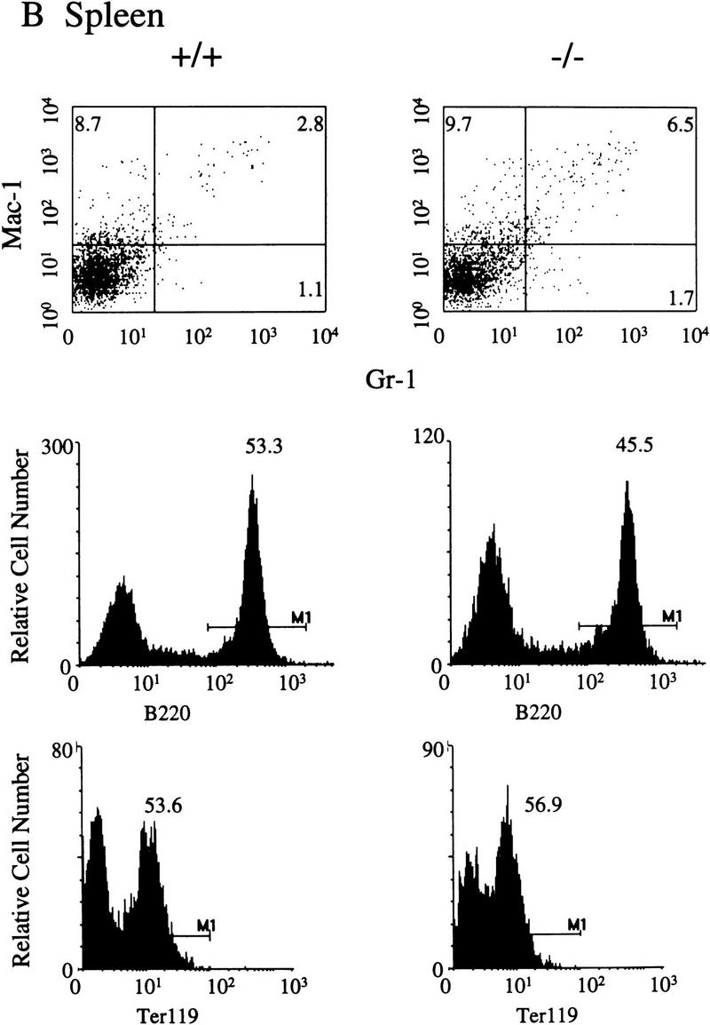
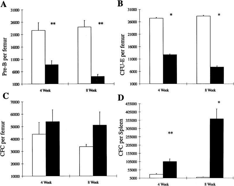
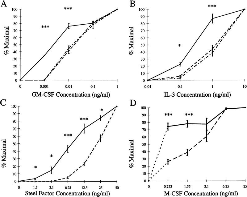
SHIP is a 145-kD SH2-containing inositol-5-phosphatase widely expressed in hemopoietic cells. It was first identified as a tyrosine phosphoprotein associated with Shc in response to numerous cytokines. SHIP has been implicated in FcgammaRIIB receptor-mediated negative signaling in B cells and mast cells and is postulated to down-regulate cytokine signal transduction in myeloid cells. To define further its role in the proliferation and differentiation of hemopoietic progenitors, as well as its function in mature cells, we have generated embryonic stem cells and mice bearing a targeted disruption of both SHIP alleles. Here we show that although SHIP null mice are viable and fertile, they fail to thrive and survival is only 40% by 14 weeks of age. Mortality is associated with extensive consolidation of the lungs resulting from infiltration by myeloid cells. Increased numbers of granulocyte-macrophage progenitors are observed in both the bone marrow and spleen of SHIP-/- mice, perhaps as a consequence of hyper-responsiveness to stimulation by macrophage-colony stimulating factor, granulocyte-macrophage colony stimulating factor, interleukin-3, or Steel factor as observed in vitro. In contrast, numbers of bone marrow lymphoid and late erythroid progenitors (CFU-E) are reduced. Thus, homozygous disruption of SHIP establishes the crucial role of this molecule in modulating cytokine signaling within the hemopoietic system and provides a powerful model for further delineating its function.


Figures

Figure 1
Figure 1
Targeted disruption of the murine SHIP gene. (A) Partial restriction map of the wild-type 129 SHIP gene (Wild type), the tk-containing targeting vector that replaces the coding sequence of the first exon with the Neo-resistance gene in the antisense orientation (Vector), and the organization of the targeted 129 allele (Targeted). (Dashed lines) Area of homology between the vector and the endogenous gene. The 2.0-kb genomic probe used for screening is indicated along with expected sizes of the wild-type and targeted KpnI fragments. (B) Genomic Southern blot analysis using a 2-kb KpnI–HindIII genomic probe. The nontargeted SHIP allele is visualized as a 4.6-kb band in the wild-type ES cells and a 2.9-kb C57Bl/6 band in the mice (because of a polymorphism at this locus), whereas the SHIP−/− cells and mice contain only the targeted 5.8-kb band. (C) Western blot analysis. R1 SHIP wild-type (+/+), heterozygous (+/−), and null (−/−) ES cells were differentiated in vitro as described previously (Helgason et al. 1996). Hemopoietic cells were derived from suspension cultures of day 10 embryoid bodies, and total cell lysates were probed with an antibody against the carboxy-terminal region spanning the two NPxY sequences of SHIP. The predominant 145- and 135-kD forms are indicated at left. Total cell lysates were also prepared from freshly isolated bone marrow of representative F2 mice and blots were probed with an antibody against the amino terminal SH2 domain. The predominant 110-kD protein is indicated at right.
Figure 1
Figure 1
Targeted disruption of the murine SHIP gene. (A) Partial restriction map of the wild-type 129 SHIP gene (Wild type), the tk-containing targeting vector that replaces the coding sequence of the first exon with the Neo-resistance gene in the antisense orientation (Vector), and the organization of the targeted 129 allele (Targeted). (Dashed lines) Area of homology between the vector and the endogenous gene. The 2.0-kb genomic probe used for screening is indicated along with expected sizes of the wild-type and targeted KpnI fragments. (B) Genomic Southern blot analysis using a 2-kb KpnI–HindIII genomic probe. The nontargeted SHIP allele is visualized as a 4.6-kb band in the wild-type ES cells and a 2.9-kb C57Bl/6 band in the mice (because of a polymorphism at this locus), whereas the SHIP−/− cells and mice contain only the targeted 5.8-kb band. (C) Western blot analysis. R1 SHIP wild-type (+/+), heterozygous (+/−), and null (−/−) ES cells were differentiated in vitro as described previously (Helgason et al. 1996). Hemopoietic cells were derived from suspension cultures of day 10 embryoid bodies, and total cell lysates were probed with an antibody against the carboxy-terminal region spanning the two NPxY sequences of SHIP. The predominant 145- and 135-kD forms are indicated at left. Total cell lysates were also prepared from freshly isolated bone marrow of representative F2 mice and blots were probed with an antibody against the amino terminal SH2 domain. The predominant 110-kD protein is indicated at right.
Figure 2
Figure 2
Decreased survival of SHIP−/− mice. F2 littermates [14 +/+ (diamonds) , 22 +/− (circles), and 14 −/− (squares)] were followed over time and the percentage of surviving mice is indicated as a function of time.
Figure 3
Figure 3
Lung pathology observed in SHIP−/− mice. (A) Representative gross appearance of lungs from a 4-week-old wild-type (left) and two 4-week-old SHIP−/− mice (right) showing diffuse enlargement and patchy pleural discoloration of the lungs from the knockout mice. (B) Low power micrograph of H&E stained sections from a representative 8-week-old SHIP−/− mouse showing patchy cellular infiltrates in alveolar airspaces adjacent to relatively normal appearing lung. (C) High power photomicrograph of H&E stained sections from an 8-week-old knockout mouse demonstrating alveolar infiltration by lipid-laden macrophages, including multinucleated forms, as well as collections of neutrophils mixed with the macrophages. (D). Chloracetate esterase stains for macrophages showing strong reactivity of alveolar cell infiltrates of the SHIP−/− lung.
Figure 4
Figure 4
Representative FACS profiles of 4–5-week-old SHIP F2 mice. Five mice of each genotype were analyzed and the average percentages of positive cells determined. A FACS profile representative of each of these values is presented for bone marrow (A), spleen (B), and thymus (C) of SHIP wild-type (+/+) or null (−/−) mice. Numbers represent the percent of viable (PI), positively stained cells within each marked region.
Figure 4
Figure 4
Representative FACS profiles of 4–5-week-old SHIP F2 mice. Five mice of each genotype were analyzed and the average percentages of positive cells determined. A FACS profile representative of each of these values is presented for bone marrow (A), spleen (B), and thymus (C) of SHIP wild-type (+/+) or null (−/−) mice. Numbers represent the percent of viable (PI), positively stained cells within each marked region.
Figure 5
Figure 5
Altered myelopoiesis, erythropoiesis, and lymphopoiesis in SHIP−/− mice. In vitro hematopoietic colony formation was examined in methylcellulose colony assays using cells from wild-type (open bars) or null (solid bars) SHIP F2 littermates at 4–5 weeks and 8–10 weeks of age. (A) Numbers of pre-B progenitors per femur detected in methylcellulose containing 10 ng/ml IL-7. (B) Numbers of day 2 CFU-E per femur detected in methylcellulose supplemented with 50 ng/ml SF and 3 U/ml Epo. Granulocyte–macrophage CFC per femur (C) or per spleen (D) were detected in methylcellulose containing 10 ng/ml IL-3, 10 ng/ml IL-6, 50 ng/ml SF, and 3 U/ml Epo. In all cases, values represent the mean ± s.e.m. of 3 or more duplicate determinations. Statistical significance compared to +/+ littermates was determined using the Student’s t-test. P ≤ 0.05 (*); P ≤ 0.01 (**).
Figure 6
Figure 6
Altered growth factor responsiveness of SHIP−/− CFC. Bone marrow derived from wild type (– –), heterozygous (– . –), or null (—) littermates was plated in methylcellulose containing the indicated concentrations of (A) GM-CSF, (B) IL-3, (C) Steel factor, or (D) M–CSF. Colonies of >20 cells were scored on day 10 of culture and the numbers of colonies present at each cytokine concentration were calculated as the percent of the number formed in the highest concentration of the cytokine indicated. Values represent the mean ± s.e.m. for duplicate determinations using at least three mice per group. Statistical significance compared to +/+ littermates was determined using the Student’s t-test. P ≤ 0.05 (*); P ≤ 0.008 (***).

Similar articles

Cited by

References

    1. Cutler RL, Liu L, Damen JE, Krystal G. Multiple cytokines induce the tyrosine phosphorylation of Shc and its association with Grb2 in hemopoietic cells. J Biol Chem. 1993;268:21463–21465. - PubMed
    1. Damen JE, Liu L, Cutler RL, Krystal G. Erythropoietin stimulates the tyrosine phosphorylation of Shc and its association with Grb2 and a 145-Kd tyrosine phosphorylated protein. Blood. 1993;82:2296–2303. - PubMed
    1. Damen JE, Liu L, Rosten P, Humphries RK, Jefferson AB, Majerus PW, Krystal G. The 145-kDa protein induced to associate with Shc by multiple cytokines is an inositol tetraphosphate and phosphatidylinositol 3,4,5-trisphosphate 5-phosphatase. Proc Natl Acad Sci. 1996;93:1689–1693. - PMC - PubMed
    1. Helgason CD, Sauvageau G, Lawrence HJ, Largman C, Humphries RK. Overexpression of HOXB4 enhances the hematopoietic potential of embryonic stem cells differentiated in vitro. Blood. 1996;87:2740–2749. - PubMed
    1. Jiao H, Yang W, Berrada K, Tabrizi M, Shultz L, Yi T. Macrophages from motheaten and viable motheaten mutant mice show increased proliferative responses to GM-CSF: Detection of potential HCP substrates in GM-CSF signal transduction. Exp Hematol. 1997;25:592–600. - PubMed

Publication types

MeSH terms

Substances