. 1998 Jun 1;12(11):1665-77.
doi: 10.1101/gad.12.11.1665.

The surveillance complex interacts with the translation release factors to enhance termination and degrade aberrant mRNAs

Affiliations

The surveillance complex interacts with the translation release factors to enhance termination and degrade aberrant mRNAs

K Czaplinski et al. Genes Dev. .

Abstract

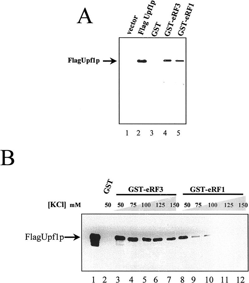
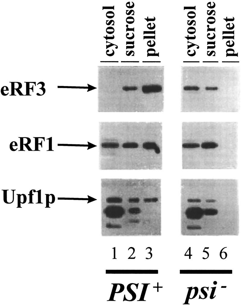
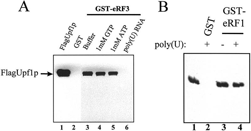
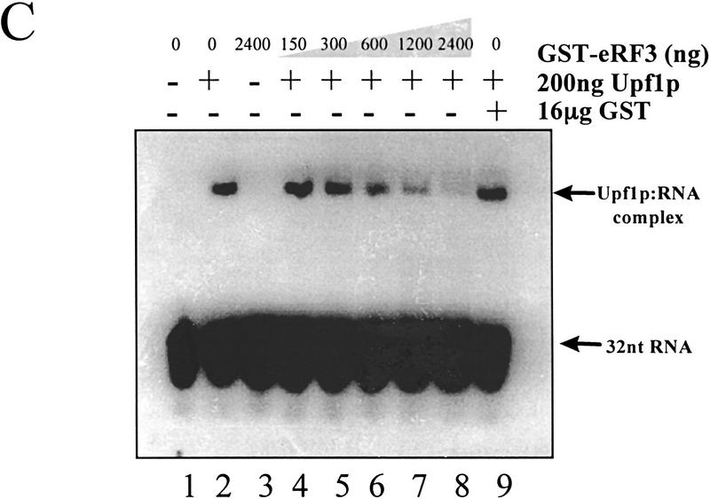
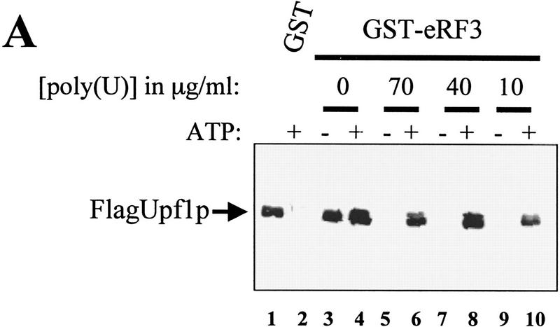
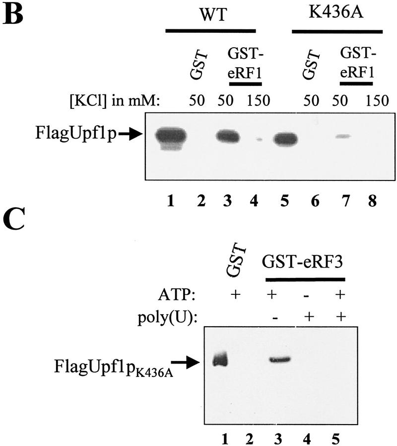
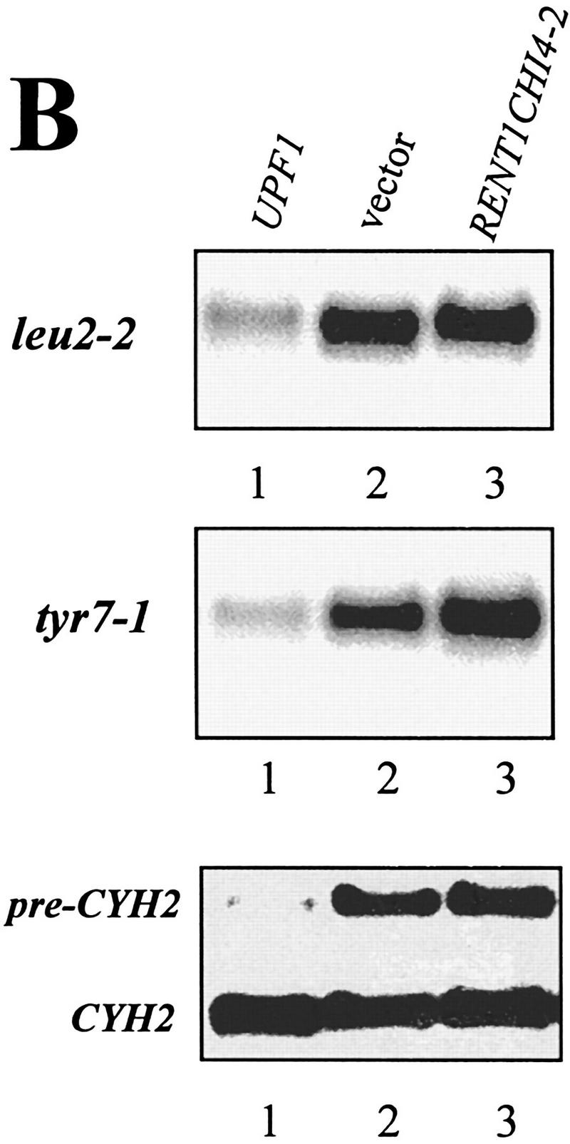
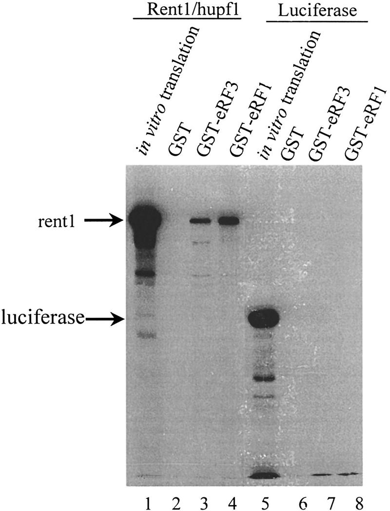
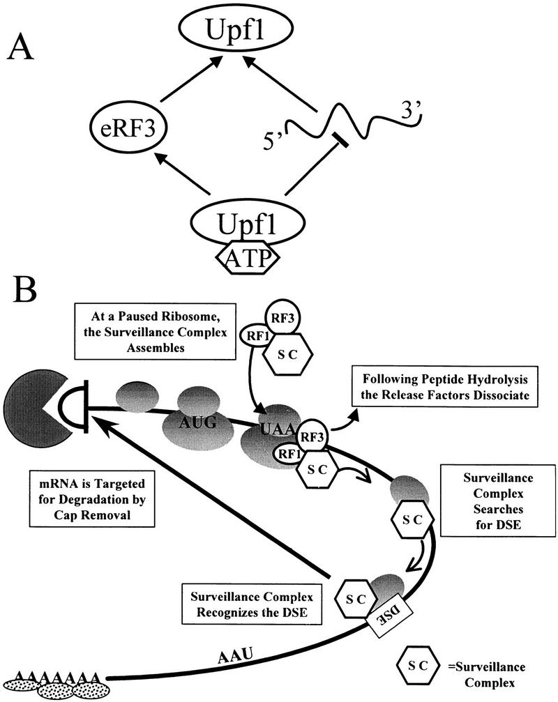
The nonsense-mediated mRNA decay pathway is an example of an evolutionarily conserved surveillance pathway that rids the cell of transcripts that contain nonsense mutations. The product of the UPF1 gene is a necessary component of the putative surveillance complex that recognizes and degrades aberrant mRNAs. Recent results indicate that the Upf1p also enhances translation termination at a nonsense codon. The results presented here demonstrate that the yeast and human forms of the Upf1p interact with both eukaryotic translation termination factors eRF1 and eRF3. Consistent with Upf1p interacting with the eRFs, the Upf1p is found in the prion-like aggregates that contain eRF1 and eRF3 observed in yeast [PSI+] strains. These results suggest that interaction of the Upf1p with the peptidyl release factors may be a key event in the assembly of the putative surveillance complex that enhances translation termination, monitors whether termination has occurred prematurely, and promotes degradation of aberrant transcripts.


Figures

Figure 1
Figure 1
The yeast Upf1 protein interacts specifically with the peptidyl release factors. (A) GST–eRF1 or GST–eRF3 fusion proteins bind specifically to Upf1p in a yeast extract. Cytoplasmic extracts from a yeast strain BJ3505 transformed with either pG-1 (vector) or pG-1FLAGUPF1 (Flag-Upf1p) were prepared in IBTB and incubated with 30 μl of GST, GST–eRF1, or GST–eRF3 Sepharose–protein complexes. The Sepharose–protein complexes were washed twice in IBTB (see Materials and Methods), resuspended in SDS–polyacrylamide loading buffer, separated on an 8% SDS-polyacrylamide gel, and immunoblotted by use of anti-Flag antibody. (B) Upf1p interacts directly with both eRF1 and eRF3. Upf1p was purified as described previously (Czaplinski et al. 1995). Upf1p (200 ng) was added to 10 μl of GST, GST–eRF1, or GST–eRF3 Sepharose–protein complexes in a total reaction volume of 200 μl in IBTB supplemented with KCl to the final concentration indicated above each lane. After 1 hr at 4°C, Sepharose–protein complexes were washed for 3 min with 1 ml of IBTB supplemented with KCl to the final concentration indicated above each lane. The purified Sepharose–protein complexes were resuspended in SDS-polyacrylamide loading buffer and separated on a 7.5% SDS-polyacrylamide gel and immunoblotted as in A.
Figure 2
Figure 2
The Upf1p is associated with eRF3 [PSI+] aggregates. Cytoplasmic extracts from isogenic [PSI+] and [psi] variants of strain 7G-H66 upf1Δ and containing FLAG–UPF1 inserted into a centromere plasmid were fractionated by centrifugation through a sucrose cushion as described previously (Paushkin et al. 1997b). Supernatant (cytosol), sucrose pad (sucrose), and pellet fractions were subjected to SDS–PAGE, and the distribution of eRF1, eRF3, and Upf1p within these fractions was determined by immunoblotting with polyclonal antibody against eRF1 and eRF3 and a monoclonal antibody against the Flag epitope. A 95-kD protein cross-reacts with anti-flag antibody in strain 7G-H66 and has the same distribution in [PSI+] and [psi] cells. This 95-kD protein is not present in extracts prepared from strain BJ3505 (see Fig. 1).
Figure 3
Figure 3
eRF3 and RNA compete for binding to Upf1p. (A) Poly(U) RNA prevents Upf1p from binding to eRF3. Reaction mixtures were prepared as described in Fig. 1B, except that binding was performed in TBSTB (TBST with 100 μg/ml BSA) and reaction mixtures contained 1 mm ATP, 1 mm GTP, or 100 μg/ml poly(U) RNA as indicated above each lane. The reaction mixtures were mixed for 1 hr at 4°C. Following mixing, the complexes were washed as in Fig. 1B with TBSTB containing 1 mm ATP, 1 mm GTP, or 100 μg/ml poly(U) RNA as indicated above each lane. (B) Poly(U) RNA does not prevent Upf1 and eRF1 interaction. Reaction mixtures were prepared as in Fig. 1B, in the presence or absence of 100 μg/ml poly(U) RNA as indicated above each lane. (C) eRF3 inhibits Upf1p RNA binding. A uniformly labeled 32-nucleotide RNA was synthesized by SP6 transcription of SstI-digested pGEM5Zf(+). The indicated amounts of GST–eRF3 were incubated with 200 ng of Upf1p for 15 min at 4°C. RNA substrate (50 fmoles) was added and incubated for 5 min. Stop solution was added, and reactions electrophoresed in a 4.5% native polyacrylamide gel (0.5× TBE, 30:0.5 acrylamide/bisacrylamide, with 5% glycerol).
Figure 3
Figure 3
eRF3 and RNA compete for binding to Upf1p. (A) Poly(U) RNA prevents Upf1p from binding to eRF3. Reaction mixtures were prepared as described in Fig. 1B, except that binding was performed in TBSTB (TBST with 100 μg/ml BSA) and reaction mixtures contained 1 mm ATP, 1 mm GTP, or 100 μg/ml poly(U) RNA as indicated above each lane. The reaction mixtures were mixed for 1 hr at 4°C. Following mixing, the complexes were washed as in Fig. 1B with TBSTB containing 1 mm ATP, 1 mm GTP, or 100 μg/ml poly(U) RNA as indicated above each lane. (B) Poly(U) RNA does not prevent Upf1 and eRF1 interaction. Reaction mixtures were prepared as in Fig. 1B, in the presence or absence of 100 μg/ml poly(U) RNA as indicated above each lane. (C) eRF3 inhibits Upf1p RNA binding. A uniformly labeled 32-nucleotide RNA was synthesized by SP6 transcription of SstI-digested pGEM5Zf(+). The indicated amounts of GST–eRF3 were incubated with 200 ng of Upf1p for 15 min at 4°C. RNA substrate (50 fmoles) was added and incubated for 5 min. Stop solution was added, and reactions electrophoresed in a 4.5% native polyacrylamide gel (0.5× TBE, 30:0.5 acrylamide/bisacrylamide, with 5% glycerol).
Figure 4
Figure 4
ATP prevents RNA from competing with eRF3 for binding to Upf1p. (A) Reaction mixtures were prepared as described in Fig. 3A, except that binding was performed in IBTB, and reaction mixtures contained 1 mm ATP or poly(U) RNA at the concentrations indicated above each lane. The reaction mixtures were mixed for 1 hr at 4°C. Following mixing, the complexes were washed with IBTB containing 1 mm ATP or poly(U) RNA at concentrations as indicated above each lane. (B) Upf1pK436A interacts weakly with eRF1. Reaction mixtures were prepared as in Fig. 1B, substituting Upf1pK436A for the wild-type protein (lanes 58). (C) A mutant Upf1p is unable to interact with eRF3 in the presence of RNA. Reaction mixtures were prepared as in A, substituting purified Upf1pK436A for the wild-type protein. Reactions contained 1 mm ATP or 40 μg/ml poly(U) RNA as indicated above each lane.
Figure 4
Figure 4
ATP prevents RNA from competing with eRF3 for binding to Upf1p. (A) Reaction mixtures were prepared as described in Fig. 3A, except that binding was performed in IBTB, and reaction mixtures contained 1 mm ATP or poly(U) RNA at the concentrations indicated above each lane. The reaction mixtures were mixed for 1 hr at 4°C. Following mixing, the complexes were washed with IBTB containing 1 mm ATP or poly(U) RNA at concentrations as indicated above each lane. (B) Upf1pK436A interacts weakly with eRF1. Reaction mixtures were prepared as in Fig. 1B, substituting Upf1pK436A for the wild-type protein (lanes 58). (C) A mutant Upf1p is unable to interact with eRF3 in the presence of RNA. Reaction mixtures were prepared as in A, substituting purified Upf1pK436A for the wild-type protein. Reactions contained 1 mm ATP or 40 μg/ml poly(U) RNA as indicated above each lane.
Figure 5
Figure 5
eRF1 and eRF3 inhibit Upf1p RNA-dependent ATPase activity. Upf1p RNA-dependent ATPase activity was determined in the presence of GST–RF fusions by a charcoal assay with 1 μg/ml poly(U) RNA and 100 μg/ml BSA. The results are plotted as picomoles of 32P released vs. the amount of the indicated protein. (○) GST; (□) GST–eRF3; (▵) GST–eRF1.
Figure 6
Figure 6
A RENT1/HUPF1 chimeric allele functions in translation termination. (A) A RENT1/HUPF1 chimeric allele prevents nonsense suppression in a upf1Δ strain. Strain PLY146 (MATα ura3-52 trp1Δ upf1::URA3 leu2-2 tyr7-1) was transformed with YCplac22 (vector), YCpUPF1 (UPF1), YCpRent1CHI4-2, or YEpRent1CHI4-2, and cells were grown to OD600 = 0.5 in -Trp -Met medium. Dilutions of 1/10, 1/100, and 1/1000 were prepared in -Trp -Met medium and 5 μl of these dilutions was plated simultaneously on -Trp -Met (top plate) or -Trp -Met -Leu -Tyr (bottom plate) media. Cells were monitored for growth at 30°C. (B) A RENT1/HUPF1 chimeric allele does not promote decay of nonsense containing mRNAs. Total RNA was isolated from cells at OD600 = 0.8 from the strains described in A. RNA (40 μg) from strains PLY146 transformed with YCplac22 (vector), YCpUPF1 (UPF1), or YEpRent1CHI4-2 (YEpRENT1CHI4-2)(10) was subjected to Northern blotting analysis and probed with either the LEU2, TYR7, or CYH2 probes.
Figure 6
Figure 6
A RENT1/HUPF1 chimeric allele functions in translation termination. (A) A RENT1/HUPF1 chimeric allele prevents nonsense suppression in a upf1Δ strain. Strain PLY146 (MATα ura3-52 trp1Δ upf1::URA3 leu2-2 tyr7-1) was transformed with YCplac22 (vector), YCpUPF1 (UPF1), YCpRent1CHI4-2, or YEpRent1CHI4-2, and cells were grown to OD600 = 0.5 in -Trp -Met medium. Dilutions of 1/10, 1/100, and 1/1000 were prepared in -Trp -Met medium and 5 μl of these dilutions was plated simultaneously on -Trp -Met (top plate) or -Trp -Met -Leu -Tyr (bottom plate) media. Cells were monitored for growth at 30°C. (B) A RENT1/HUPF1 chimeric allele does not promote decay of nonsense containing mRNAs. Total RNA was isolated from cells at OD600 = 0.8 from the strains described in A. RNA (40 μg) from strains PLY146 transformed with YCplac22 (vector), YCpUPF1 (UPF1), or YEpRent1CHI4-2 (YEpRENT1CHI4-2)(10) was subjected to Northern blotting analysis and probed with either the LEU2, TYR7, or CYH2 probes.
Figure 7
Figure 7
Rent1/hupf1 interacts with eRF1 and eRF3. NotI linearized pT7RENT1 (lanes 14) or luciferase template (lanes 58) was used in the TNT-coupled reticulocyte in vitro transcription translation as per manufacturer’s directions (Promega). Completed translation reactions (2 μl) were electrophoresed in lanes 1 and 5. Completed reactions (5 μl) were incubated in 200 μl of IBTB with 10 μl of GST, GST–eRF1, or GST–eRF3 Sepharose–protein complexes as indicated above each lane. Following mixing for 1 hr at 4°C, the Sepharose–protein complexes were washed as in Fig. 1A, and the bound proteins were subjected to SDS-PAGE in an 8% gel. Following electrophoresis, gels were fixed for 30 min in 50% methanol, 10% acetic acid, and then treated with 1 m salicylic acid for 1 hr. Gels were dried and subjected to autoradiography.
Figure 8
Figure 8
Model for Upf1 function in mRNA surveillance. (A) Modulation of RNA binding enhances interaction of Upf1 with peptidyl release factors. ATP binding to Upf1p decreases the affinity of Upf1 for RNA. Because RNA and eRF3 compete for binding to Upf1, interaction with eRF3 is favored. (B) A model for mRNA surveillance. Interaction of Upf1p with peptidyl release factors assembles an mRNA surveillance complex at a termination event. This interaction prevents Upf1 from binding RNA and hydrolyzing ATP, and enhances translation termination. Following peptide hydrolysis, the release factors dissociate from the ribosome, activating the Upf1p helicase activity. The surveillance complex then scans 3′ of the termination codon for a DSE. Interaction of the surveillance complex with the DSE signals that premature translation termination has occurred and the mRNA is then decapped and degraded by the Dcp1p and Xrn1p exoribonuclease, respectively.

Similar articles

Cited by

References

    1. Altamura N, Groudinsky O, Dujardin G, Slonimski PP. NAM7 nuclear gene encodes a novel member of a family of helicases with a Z1-6n-ligand motif and is involved in mitochondrial functions in Saccharomyces cerevisiae. J Mol Biol. 1992;224:575–587. - PubMed
    1. Andjelkovic N, Zolnierowicz S, Van Hoof C, Goris J, Hemmings BA. The catalytic subunit of protein phosphatase 2A associates with the translation termination factor eRF1. EMBO J. 1996;15:7156–7167. - PMC - PubMed
    1. Applequist SE, Selg M, Roman C, Jack HM. Cloning and characterization of HUPF1, a human homolog of the Saccharomyces cerevisiae nonsense mRNA-reducing UPF1 protein. Nucleic Acids Res. 1997;25:814–821. - PMC - PubMed
    1. Atkin AL, Altamura N, Leeds P, Culbertson MR. The majority of yeast UPF1 co-localizes with polyribosomes in the cytoplasm. Mol Biol Cell. 1995;6:611–625. - PMC - PubMed
    1. Atkin AL, Schenkman LR, Eastham M, Dahlseid JN, Lelivelt MJ, Culbertson MR. Relationship between yeast polyribosomes and Upf proteins required for nonsense mediated mRNA decay. J Biol Chem. 1997;272:22163–22172. - PubMed

Publication types