. 1998 Jun 1;12(11):1691-704.
doi: 10.1101/gad.12.11.1691.

Multistep signaling requirements for pituitary organogenesis in vivo

Affiliations

Multistep signaling requirements for pituitary organogenesis in vivo

M Treier et al. Genes Dev. .

Abstract

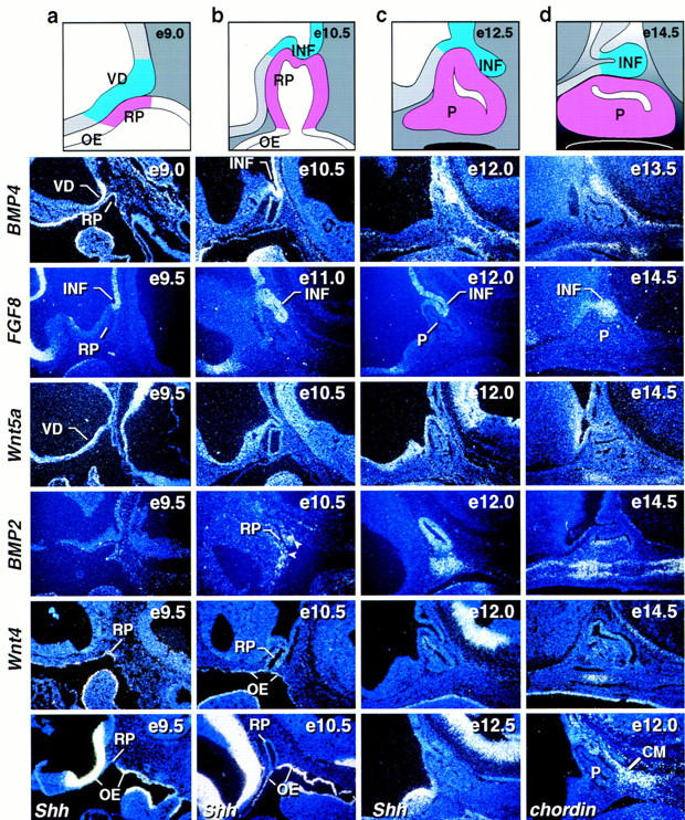
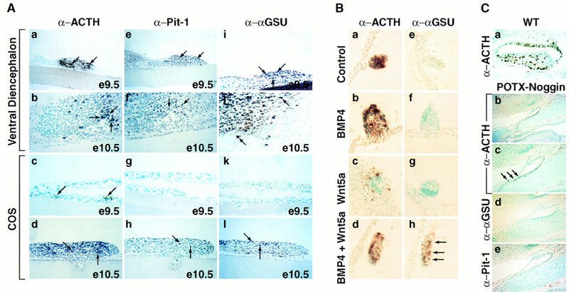
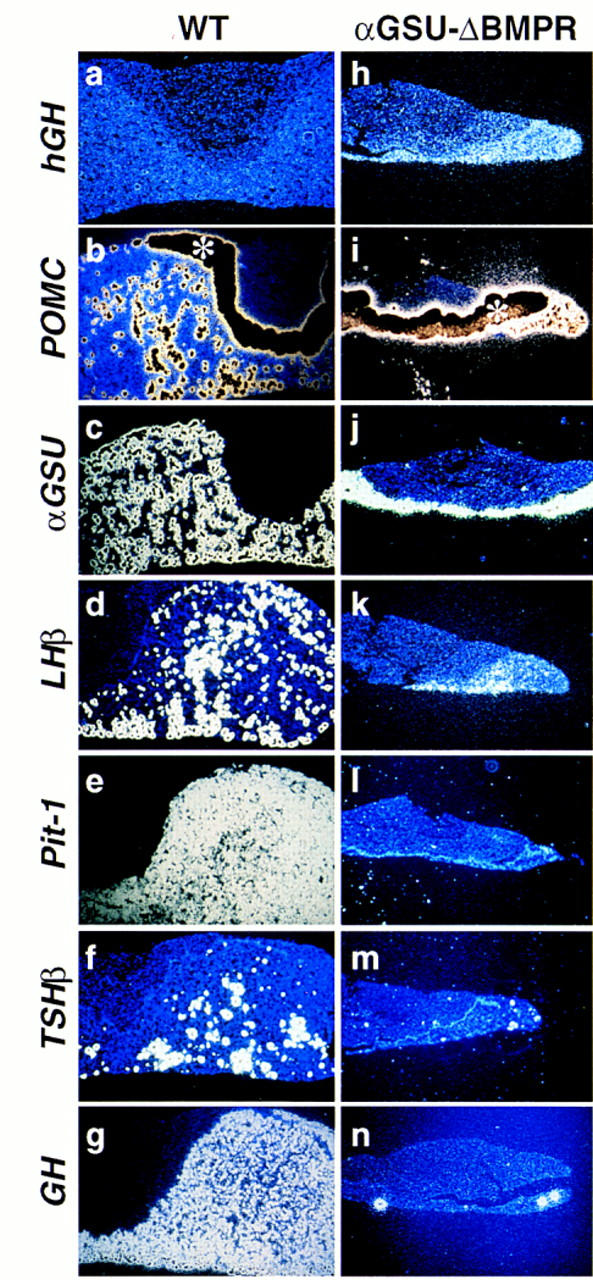
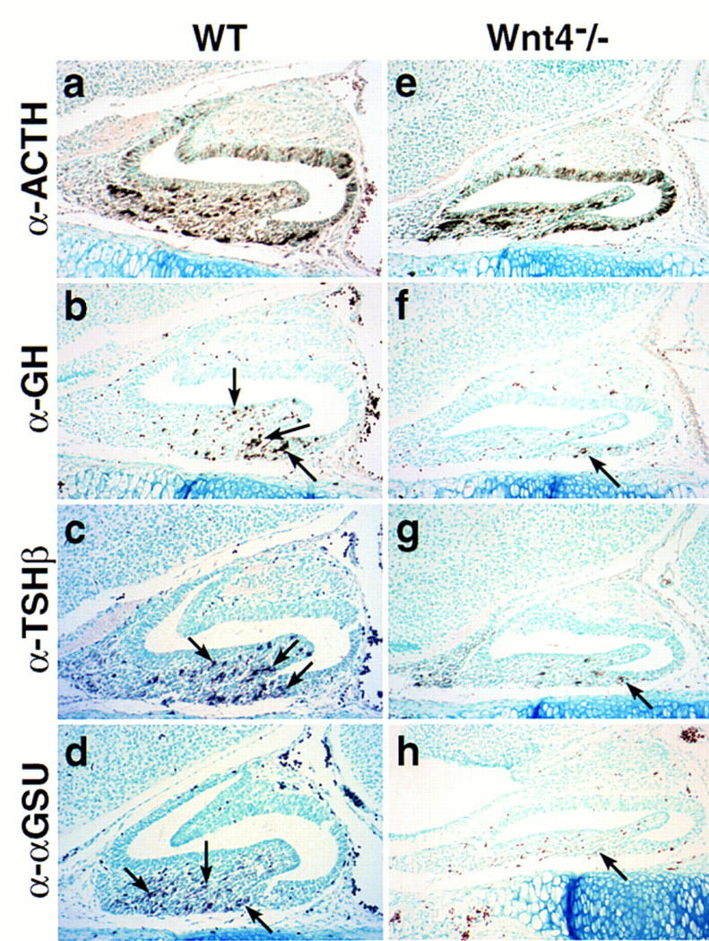
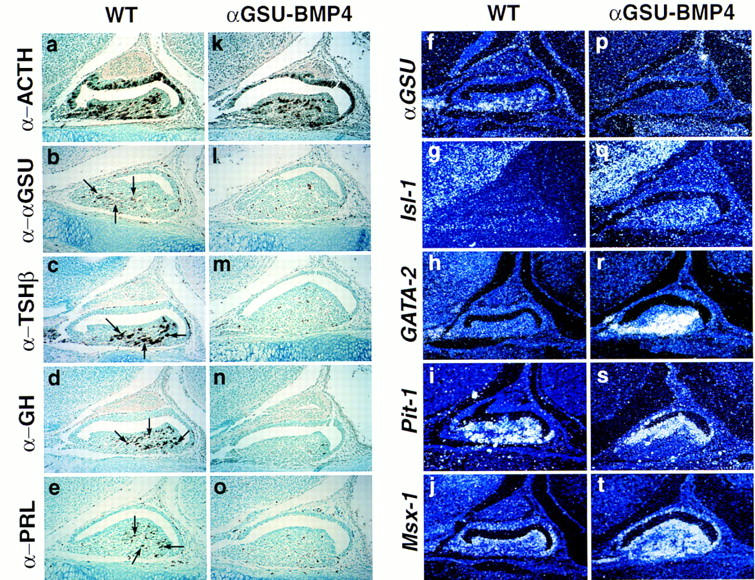
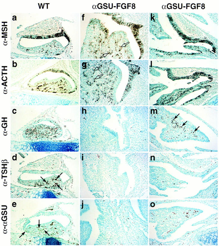
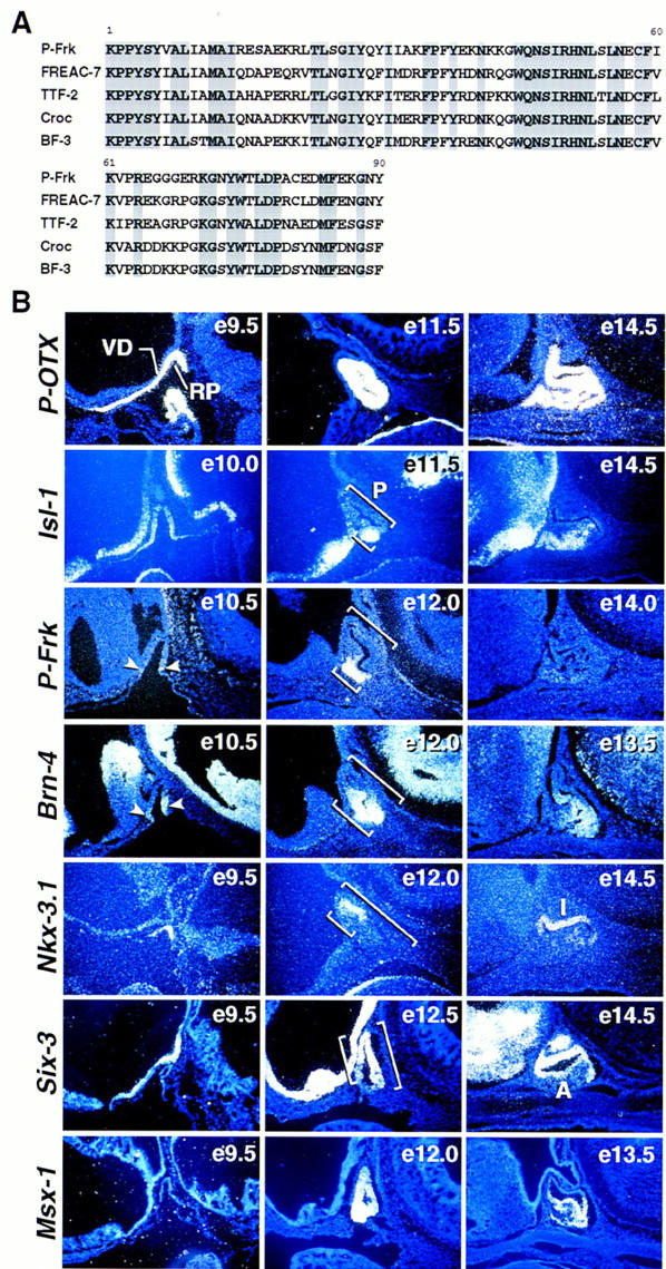
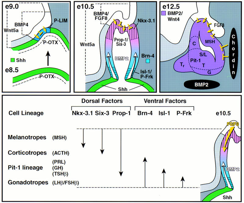
During development of the mammalian pituitary gland specific hormone-producing cell types, critical in maintaining homeostasis, emerge in a spatially and temporally specific fashion from an ectodermal primordium. We have investigated the molecular basis of generating diverse pituitary cell phenotypes from a common precursor, providing in vivo and in vitro evidence that their development involves three sequential phases of signaling events and the action of a gradient at an ectodermal boundary. In the first phase, the BMP4 signal from the ventral diencephalon, expressing BMP4, Wnt5a, and FGF8, represents a critical dorsal neuroepithelial signal for pituitary organ commitment in vivo. Subsequently, a BMP2 signal emanates from a ventral pituitary organizing center that forms at the boundary of a region of oral ectoderm in which Shh expression is selectively excluded. This BMP2 signal together with a dorsal FGF8 signal, appears to create opposing activity gradients that are suggested to generate overlapping patterns of specific transcription factors underlying cell lineage specification events, whereas Wnt4 is needed for the expansion of ventral pituitary cell phenotypes. In the third phase, temporally specific loss of the BMP2 signal is required to allow terminal differentiation. The consequence of these sequential organ and cellular determination events is that each of the hormone-producing pituitary cell types-gonadotropes, thyrotropes, somatotropes, lactotropes, corticotropes, and melanotropes-appear to be determined, in a ventral-to-dorsal gradient, respectively.


Figures

Figure 1
Figure 1
Expression of signaling molecules during early stages of pituitary development. Schematic representation of different stages of pituitary development (a–d). In situ hybridization analysis for BMP4, FGF8, Wnt5a, BMP2, Wnt4, Sonic hedgehog (Shh), and chordin during early stages of pituitary development [embryonic day (e) of mouse development is indicated at top right of each panel]. BMP4 expression is detected early on in the ventral diencephalon and later in the infundibulum to subsequently disappear. FGF8 is expressed temporally later than BMP4 in the infundibulum. Wnt5a is expressed throughout the ventral diencephalon not showing any restriction. No BMP2 expression is detected early in the vicinity of Rathke’s pouch. At E10.5 BMP2 expression was detected in the most ventral part of Rathke’s pouch ectoderm and in some adjacent mesenchymal cells (arrows). By E12.0 BMP2 was expressed throughout Rathke’s pouch and in the underlying condensing mesenchymal cells. Two days later BMP2 expression is confined to the perilumenal cells in Rathke’s pouch and the underlying cartilage growth zone. Wnt4 expression is observed in the oral ectoderm and Rathke’s pouch throughout pituitary gland development. Shh expression is observed throughout the oral ectoderm, whereas the invaginating part of oral ectoderm that becomes Rathke’s pouch is void of any detectable expression, creating a molecular boundary between two ectodermal domains of Shh expressing and nonexpressing cells. The BMP antagonist chordin is expressed in the caudal mesenchyme (CM) adjacent to Rathke’s pouch. BMP2 and Wnt4 expression is noted in the condensing mesenchyme beginning at E12. (INF) infundibulum; (RP) Rathke’s pouch; (OE) oral ectoderm; (P) pituitary; (VD) ventral diencephalon.
Figure 2
Figure 2
BMP activity is required for anterior pituitary development. (A) Requirement for signaling from the ventral diencephalon in the induction of pituitary lineages. (a,e,i) Coculturing of E9.5 ventral diencephalon with E9.5 Rathke’s pouch explant for 6 days resulted in the appearance of all major lineages as shown by immunohistochemical staining with ACTH (a), Pit-1 (e), and αGSU (i) antisera. (b,f,j) Coculturing experiments done under the same conditions with explants from E10.5, respectively, resulted in the same outcome as in a,e, and i). (c,g,k) Coculturing E9.5 Rathke’s pouch explants in the presence of COS cells for 6 days led to the appearance of only few ACTH-positive cells (arrows, c), no Pit-1 and αGSU-positive cells were detected (g,k). (d,h,l) Coculturing experiment done under the same conditions as under (c,g,and k) with Rathke’s pouch explants from E10.5, resulting in the appearance of ACTH (d)-, Pit-1 (h)-, and αGSU (l)-positive cells. Results are shown from n = 5 independent experiments. (B) Synergistic induction of αGSU expression through BMP and Wnt signaling. (a,e) E9.5 Rathke’s pouch explants cultured in the presence of COS cells showed induction of only ACTH expression (a) as revealed by immunohistochemistry; no αGSU expression (e) is detectable. (b,c,f,g) Similar results were obtained as in a and e when E9.5 Rathke’s pouch explants were cultured with COS cells secreting either BMP4 or Wnt5a alone. (d,h) Coculturing of COS cells producing both BMP4 and Wnt5a with E9.5 Rathke’s pouch explants resulted in induction of αGSU expression (h). Results are from n = 4 independent experiments. (C) Expression of a POTX–Noggin transgene leads to an arrest of pituitary development. (a) Wild-type (WT) littermate at E17.5 stained for ACTH. (b) Phenotypic appearance of the pituitary gland in a POTX-Noggin transgenic embryo at E17.5. The gland has not progressed beyond a single layer epithelium stage reminiscent of Rathke’s pouch at E10.0. A connection towards the oral cavity still remains. (c) Higher magnification of b. Arrows mark the few ACTH-positive cells that can be found in this gland. (d,e) Immunohistochemical staining of adjacent sections with αGSU and Pit-1 antisera, respectively. No positive cells for either of the two markers could be found. Four independent transgene-positive embryos from this stage of gestation were obtained with three showing an abnormal phenotype.
Figure 3
Figure 3
Role of endogenous BMP activity in cell-type determination. Pituitary phenotype of mice expressing the αGSU–ΔBMPR transgene. Pituitaries of a 8-week-old αGSU–ΔBMPR transgenic mouse (h–n) and its wild-type littermate (a–g) are shown. In situ analysis for Pit-1 (e,l) and the trophic hormones POMC (b,i), αGSU (c,j), LH (d,k), TSHβ (f,m) and GH (g,n), as well as a transgene specific probe hGH (a,h) are shown. The asterisk (*) in b marks the intermediate lobe that expresses POMC at such a high level that the region is overexposed and hence black. Three independent founders exhibited a dwarf phenotype.
Figure 4
Figure 4
Wnt4 affects cell type expansion in the pituitary gland. Pituitary phenotype in Wnt4−/− mutant animals at E17.5 (e–h) compared to wild-type littermate (a–d). Immunohistochemical analysis for the trophic hormones ACTH (a,e), GH (b,f), TSHβ (c,g), and αGSU (d,h) are shown. Arrows indicate positive cells for the corresponding hormones in the mutant and wild-type pituitaries. Only a few positive cells for GH, TSHβ, and αGSU are present in the pituitaries of Wnt4−/− mutant animals. Five pairs of embryos were analyzed.
Figure 5
Figure 5
Prolonged BMP activity inhibits terminal differentiation. Phenotypic appearance of the pituitary gland in αGSU–BMP4 transgenic embryos at E17.0. Pituitaries of an E17 transgenic embryo (k–t) and its wild-type littermate (a–j) are shown. Immunohistochemical analysis for the trophic hormones ACTH (a,k), αGSU (b,l), TSHβ (c,m), GH (d,n), and PRL (e,o) are shown. Arrows indicate positive cells for the corresponding hormones in the wild-type mouse. In situ analysis was performed with the following markers: αGSU (f,p), Isl-1 (g,q), GATA-2 (h,r), Pit-1 (i,s), and Msx-1 (j,t). Four independent transgenic embryos from this stage of gestation were analyzed, all showing an abnormal phenotype.
Figure 6
Figure 6
FGF8 promotes proliferation and opposes BMP activity in the pituitary gland. Phenotypic appearance of the pituitary gland in αGSU–FGF8 transgenic embryos at E17.5. Two transgenic embryos are shown, because the phenotype became more severe with higher copy number of the transgene [αGSU–FGF8 (f–j) strong phenotype, αGSU–FGF8 (k–o) weaker phenotype]. In both cases the gland exhibited striking dysmorphogenesis and increased cellularity (all photomicrographs taken at the same magnification). In αGSU–FGF8 (f,g), new clusters of lumenal-like cells have budded off (open arrows). Immunohistochemical analysis for the trophic hormones MSH (a,f,k), ACTH (b,g,l), GH (c,h,m), TSHβ (d,i,n), and αGSU (e,j,o) are shown, with selective loss of GH (h), TSHβ (i), and αGSU (j) in the case of the more severe phenotype and TSHβ (n) and αGSU (o) in the case of the weaker phenotype. The arrows mark clusters of positive cells for the respective hormones analyzed. Note that the dark dots, especially seen in the α–αGSU analysis of the αGSU–FGF8 (o) transgene, represent blood cells and not positive staining. Seven independent transgenic embryos at this stage were analyzed, with three showing the more severe abnormal phenotype then the other four.
Figure 7
Figure 7
Patterning of Rathke’s pouch by spatially restricted expression of transcription factors. (A) Comparison of the amino acid sequence of the new Winged-helix transcription factor P-Frk to that of other related Winged-helix (Forkhead) factors. (B) In situ hybridization analysis for P-OTX, Isl-1, P-Frk, Brn-4, Nkx-3.1, Six-3, and Msx-1 during early stages of pituitary development. Notice that P-Frk expression was detected at E10.5 in the most ventral part of Rathke’s pouch (arrowheads). Brackets mark the position of the pituitary and the different dorsal-to-ventral expression of each transcription factor, respectively. (e) Embryonic day of mouse development; (A) anterior lobe; (I) intermediate lobe; (RP) Rathke’s pouch; (P) pituitary; (VD) ventral diencephalon.
Figure 8
Figure 8
Model of cell lineage determination in pituitary ontogeny. At E8.5 (e8.5) in mouse development, Shh and P-OTX are continuously expressed throughout the oral ectoderm. Signals from the ventral diencephalon, most prominently BMP4, suppress the expression of Shh, creating a Shh-nonexpressing zone, the primordium of the nascent Rathke’s pouch and at the same time induce P-Lim/Lhx3. The boundary between the oral ectoderm expressing Shh and the nonexpressing Rathke’s pouch function as an organizing center for ventral gene induction, including BMP2. At E10.5 Rathke’s pouch has formed and the infundibulum is visible. FGF8, expressed in the infundibulum, functions antagonistically to BMP2, resulting in ventrodorsal BMP and dorsoventral FGF activity gradients in Rathke’s pouch. This leads to the induction of several temporally and spatially restricted transcription factors, postulated to combinatorially divide Rathke’s pouch into zones with different identities. These zones are proposed to impose the determination of cell lineages at this developmental stage. During the next 2 days of cell lineage expansion, the BMP2 activity gradient in Rathke’s pouch is reinforced by ventral expression of BMP2 in the underlying condensing mesenchyme and counteracted by dorsal expression of FGF8 and caudal expression of chordin.

Similar articles

Cited by

References

    1. Bach I, Rhodes SJ, Pearse RV, Heinzel T, Gloss B, Scully KM, Sawchenko PE, Rosenfeld MG. P-Lim, a LIM homeodomain factor, is expressed during pituitary organ and cell commitment and synergizes with Pit-1. Proc Natl Acad Sci. 1995;92:2720–2724. - PMC - PubMed
    1. Basler K, Struhl G. Compartment boundaries and the control of Drosophila limb pattern by hedgehog protein. Nature. 1994;368:208–214. - PubMed
    1. Bellusci S, Henderson R, Winnier G, Oikawa T, Hogan BL. Evidence from normal expression and targeted misexpression that bone morphogenetic protein (Bmp-4) plays a role in mouse embryonic lung morphogenesis. Development. 1996;122:1693–1702. - PubMed
    1. Bellusci S, Furuta Y, Rush MG, Henderson R, Winnier G, Hogan BL. Involvement of Sonic hedgehog (Shh) in mouse embryonic lung growth and morphogenesis. Development. 1997;124:53–63. - PubMed
    1. Bieberich C, Fujita K, He W, Jay G. Prostate-specific and androgen-dependent expression of a novel homeobox gene. J Biol Chem. 1996;271:31779–31782. - PubMed

Publication types

MeSH terms

Substances

Associated data